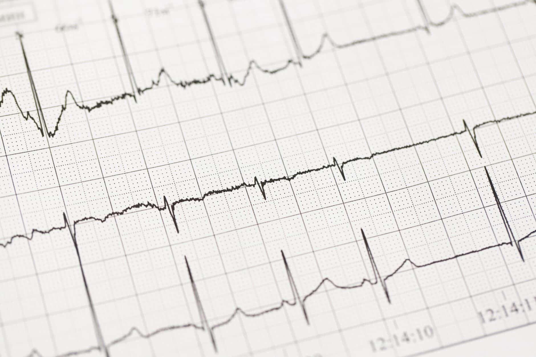
Learning to read EKGs comes naturally to some and can be more difficult for others. Typically, all it takes to get past the learning curve with EKG interpretation is the right instruction and plenty of practice.
One great way to practice your interpretation skills is to train your spot identification of different rhythms with practice EKG strips. Making your own practice rhythm strip flashcards is a great exercise in and of itself due to all of the research required. Here are some of the more important details to consider when getting started.
What Are EKG Rhythm Strips?
EKG strips are a vital tool in the diagnosis and monitoring of cardiac illnesses. They provide information on the electrical activity of the heart, showing things like beats per minute and any irregularities such as atrial fibrillation, bradycardia, and other cardiac ailments.
This data can be used to identify potential issues, confirm diagnoses, track treatment progress, and also to determine a patient’s response to medications or other interventions. Reading EKG strips accurately requires knowledge of cardiac physiology and practice interpreting them correctly. The more familiar you become with EKG strips, the better your ability will be for identifying even subtle changes that may indicate an underlying issue.
Components Of An EKG Rhythm Strip: Waves, Intervals, And Complexes
The waves on an EKG strip represent the electrical impulses that travel through the heart muscle during each heartbeat. They include P-waves (atrial depolarization), QRS-complexes (ventricular depolarization), and T-waves (ventricular repolarization). Each wave’s distinct shape can help identify arrhythmias or other heart rhythm abnormalities.
The intervals on an EKG strip measure how long certain parts of the heartbeat cycle take. For example, a PR interval measures how long it takes for atrial impulses to reach the ventricles, which helps determine if there’s any conduction blockage between these two areas of the heart muscle. The QT interval is measured to evaluate the duration of ventricular repolarization. It helps detect any prolongation that could signify an elevated hazard of sudden demise due to cardiac arrest or arrhythmia.
Complexes on an EKG strip refer to a combination of waves that make up one full heartbeat cycle, including both atrial and ventricular activities. This provides insight into the overall functioning within all four chambers of the heart simultaneously, rather than just looking at individual components separately. Examining these complexes makes it possible to detect abnormalities such as ST elevation or depression indicating myocardial infarction (heart attack) and possible coronary artery disease (CAD).
Interpreting EKG Rhythm Strips
ECG tracing is a graphical representation of the electrical activity in the heart, which can provide valuable information about its function and condition. This information is essential for healthcare providers to interpret correctly in order to diagnose and treat conditions accurately. The waveforms on an ECG tracing represent different components of the heartbeat, from atrial depolarization (P-wave) through ventricular repolarization (T-wave).
Each waveform has a distinct shape and size that indicates what type of cardiac event it represents, such as normal sinus rhythm or atrial fibrillation. With regular practice reading EKG strips, healthcare providers can improve their ability to recognize abnormal rhythms quickly and accurately so they can provide proper diagnoses and treatment plans according to their patient’s needs.
Analyzing Waveforms
To understand abnormalities accurately, it’s integral that healthcare providers understand typical waveforms. The ECG tracing consists of several waveforms representing different parts of the heartbeat. Each waveform has a specific meaning and provides information about what’s happening in your heart at any given moment.
The P-wave represents atrial depolarization, which occurs when the top chambers of your heart contract to push blood into the ventricles below them. The QRS complex represents ventricular depolarization, which occurs when both lower chambers contract to pump blood into circulation throughout your body.
Finally, the T-wave represents ventricular repolarization, which happens after both lower chambers relax to fill up with more blood for another contraction cycle to begin again soon afterward.
Identifying Abnormalities
When analyzing an ECG tracing, healthcare professionals look for abnormalities in these waveforms that could indicate underlying issues with cardiac health, such as arrhythmias or other conditions like coronary artery disease or congestive heart failure.
These abnormalities may include:
- Changes in the shape or size of one or more waves
- Prolonged pauses between beats
- Extra, skipped, or premature beats
- Inverted waves
- Wide QRS complexes indicating possible bundle branch blockage
- ST-segment elevation showing possible myocardial infarction (heart attack)
- T-wave inversion indicates possible ischemia (lack of oxygen)
Must-Include EKG Rhythms
EKG readings are an important tool for healthcare professionals to diagnose and monitor the health of their patients. Doctors and other healthcare providers can identify abnormalities in a patient’s heart rhythm by analyzing the following main reading types.
Normal Sinus Rhythm
A normal sinus rhythm is the most common type of EKG reading. It signifies that the heart’s electrical system is functioning as expected and there are no signs of cardiac issues.
In lead 2, the P wave should be upright (positive) and regular with a rate between 60-100 beats per minute (bpm). The QRS complex should also be upright (positive) with a duration of less than 0.12 seconds, follow each P wave, and have a rate between 60-100 bpm.
Atrial Fibrillation
Atrial fibrillation is when the atria have disorganized electrical activity, resulting in them trembling instead of contracting as usual. It occurs when chaotic electrical signals come from multiple sites within the atria instead of just one site. This causes an irregular heartbeat, leading to other complications such as stroke or congestive heart failure if left untreated for too long.
On an EKG strip, this will appear as a squiggly baseline representing the hundreds of P wave impulse being generated. You will also see normal, but fast, irregularly spaced QRS complexes. The QRS complexes typically beat at greater than 100 bpm.
Supraventricular Tachycardia (SVT)
Supraventricular tachycardia (SVT) is a heart condition in which the heart’s upper chambers beat abnormally quickly, leading to ventricle contractions at a rate typically between 120 and 200 bpm. SVT often has narrow QRS complexes but may also have wide ones if associated with bundle branch blockage or aberrant conduction pathways within ventricles themselves.
On an EKG strip, SVT will appear as three distinct features:
- First – a regular R-R interval, indicating that all beats originate from the same source
- Second – the presence or absence of P waves, depending upon the origin site
- Third – very narrow QRS complexes (<0.10 secs), indicating that ventricular depolarization has occurred rapidly via single focus only
Ventricular Tachycardia (VT)
Ventricular tachycardia occurs when abnormal electrical signals arise from within one or more areas inside lower chambers, causing them to contract faster than the average rate (>100 bpm). This arrhythmia usually requires immediate medical attention since prolonged episodes may eventually progress into life-threatening arrhythmias such as VFib if left untreated for too long.
On an EKG strip, this appears as wide complexes regularly occurring at rates over 100 bpm but usually below 200 bpm with inverted P waves following each QRS complex – indicative of retrograde conduction from the ventricles to the atria rather than originating within it like regular sinus rhythms do.
Tips For Practicing Rhythm Strip Interpretation
Once you have a solid understanding of the components of an EKG strip and can recognize waveforms and rhythms, you should begin to practice your interpretation skills. One way to do this is by trying out different scenarios with strips that are already labeled so you can compare your interpretations with the correct answers.
Create Practice EKG Strips Flashcards
Flashcards can be extremely helpful for both quickly referencing and taking part in extended study sessions. They are great for learning and memorizing the key terms related to ECGs, such as P waves, QRS complexes, T waves, U waves, etc.
In addition to reviewing these key terms on flashcards, practicing drawing waveforms is a great way to improve your interpretation skills more rapidly. This can be done by drawing different waveforms on one side of the flashcard so that you can easily practice and compare them with real-world examples. With enough practice and dedication, your EKG interpretation skills will quickly improve!
Practice with A Partner Or Study Group
Studying with a partner or group can also be beneficial when practicing EKG interpretation skills. Working together allows you to discuss questions and compare notes which can help deepen your understanding of the material faster than if you were studying alone.
It’s also a great way to stay motivated since having someone else hold you accountable will keep you from procrastinating or getting distracted during study sessions.
Create A Reference Guide
Creating a reference guide is another helpful strategy for mastering EKG interpretation skills. Put together all the information you need in one place, including diagrams of different waveforms, common arrhythmias, and key terms, so it’s easy for you to access whenever needed.
A reference guide shouldn’t be the end-all teaching guide for those learning EKG interpretation. It’s more of a study guide to help with quick content review.
Online Practice ECG Courses
Enrolling in an online practice ECG course can also be beneficial when learning to read electrocardiograms correctly and accurately diagnose any abnormalities in patient data sets. Online simulations provide real-life examples which allow learners to gain experience interpreting actual patient data while providing feedback along the way.
Online practice courses are invaluable for anyone looking to master their EKG reading skills quickly.
Request Our Free Practice EKG Strips Package
Using practice EKG strips is an excellent way to quickly improve your ECG interpretation skills. To help with your EKG interpretation practice, we’ve put together 10 practice EKG strips that you can download for free. Click here to get your copy now.
If you’re looking for more ways to practice your EKG interpretation, ECGEDU’s self-paced video courses may be exactly what you’re looking for. Our videos provide interactive and easy-to-follow instruction that make electrocardiogram interpretation simple and understandable. Additionally, by creatinga free account, you gain access to ECG-at-a-Glance, a one page guideline to precise ECG reading. Click here to browse our available ECG interpretation courses now.


