Introduction to Videos (AEI-W1)
This brief video describes the purpose and goals of this course.
Electrocardiogram (ECG) Basics (AEI-W1)
This chapter reviews the basics of electrocardiography. Topics include calibrations, waveforms, intervals, basic conduction, and the ECG layout.
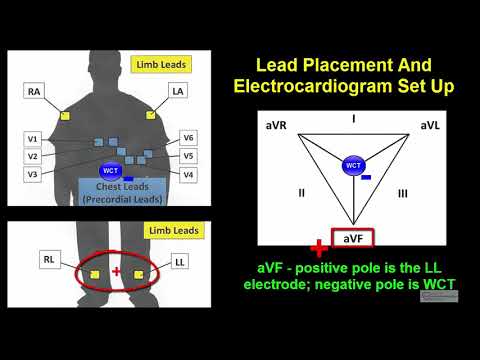
Lead Placement (AEI-W1)
This chapter reviews ECG lead placement and setup. Topics include bipolar leads, unipolar leads, lead position, right-sided leads, posterior leads, the Lewis lead, transesophageal leads, modified chest leads (MCL), and other lead configurations.
Example of Proper 12-Lead ECG Electrode Placement (AEI-W1)
This chapter gives a demonstration of proper 12-lead ECG electrode placement. It includes both the limb leads and chest leads.
Axis Determination (AEI-W1)
This chapter reviews normal and abnormal axes as well as two methods for calculating the axis. This includes the P-wave axis, the QRS axis and the T-wave axis.
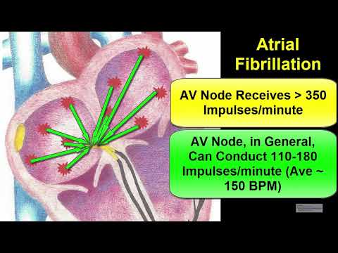
Atrial abnormalities (AEI-W1)
This chapter reviews the ECG criteria for left atrial abnormality, right atrial abnormality and biatrial abnormality.
Ventricular Hypertrophy (AEI-W1)
This chapter reviews the ECG criteria for left ventricular hypertrophy (LVH), right ventricular hypertrophy (RVH) and biventricular hypertrophy (BVH).
Bundle Branch Blocks and Intraventricular Conduction Delays (AEI-W1)
This chapter reviews the ECG criteria for right and left bundle branch blocks, non-specific intraventricular conduction delays, and incomplete right and left bundle branch blocks. Part 1 of this chapter suggests a simple way to memorize the criteria; part 2 describes an analogy to help more fully understand why these changes are seen on the ECG.
Fascicular Blocks (AEI-W1)
This chapter reviews the ECG criteria for left anterior and posterior fascicular blocks.
Ischemia, Injury, Infarction Part 1 (General findings) (AEI-W1)
This chapter reviews the ECG criteria for myocardial ischemia, injury and infarction. Part 1 details the general findings on the ECG; Part 2 details why the electrocardiogram machine sees these particular changes.
Ischemia, Injury, Infarction Part 2 (Pathophysiology) (AEI-W1)
This chapter reviews the ECG criteria for myocardial ischemia, injury and infarction. Part 1 details the general findings on the ECG; Part 2 details why the electrocardiogram machine sees these particular changes.
ST and T wave Changes (AEI-W1)
This chapter reviews primary and secondary ST and T wave changes. It is divided into two sections. Part 1 goes over the characteristic ECG findings of ST and T wave changes; and Part 2 reviews myocardial physiology and how the ECG machine see ST and T wave changes.
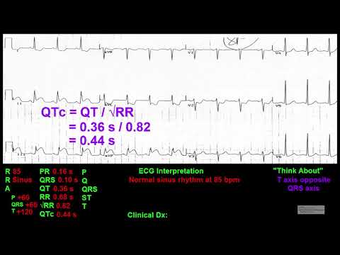
QT Interval (AEI-W1)
This chapter reviews the QT Interval. Topics include correctly measuring the QT interval, correcting the QT interval for the heart rate (QTc), and causes of prolonged and short QT intervals.
Pericarditis (AEI-W1)
This chapter reviews the ECG changes associated with pericarditis.
Central Nervous System Effects on the ECG (AEI-W1)
This chapter reviews the ECG changes seen with central nervous system abnormalities (in particular, intracranial hemorrhages).
Dextrocardia (AEI-W1)
This chapter reviews the ECG changes associated with dextrocardia.
Electrolyte Abnormalities (AEI-W1)
This chapter reviews the ECG criteria for electrolyte abnormalities including hyperkalemia, hypokalemia, hypercalcemia, and hypocalcemia. Sodium and magnesium abnormalities are touched upon.
Drug Effects (AEI-W1)
This chapter reviews ECG changes associated with digoxin, and antiarrhythmic agents. It also reviews medications that can prolong the QT interval.
Hypothermia (AEI-W1)
This chapter reviews the ECG changes associated with hypothermia.
Preexcitation and Wolff Parkinson White [WPW] (AEI-W1)
This chapter reviews ECG changes associated with preexcitation, Wolff Parkinson White (WPW Syndrome, Atrioventricular Reentrant Tachyarrhythmias (AVRT), and Atrioventricular Nodal-Type Bypass Tracts.
Lead Reversal (AEI-W1)
This chapter reviews the ECG characteristics of lead reversal. This includes: arm lead reversal and arm/leg lead reversal.
Transition (AEI-W1)
This chapter reviews transition. This includes the definitions and causes of normal transition, early transition and late transition.
Poor R Wave Progression (PRWP) (AEI-W1)
This chapter reviews poor R wave progression and R wave reversal. It goes through an algorithm that can be used to determine the cause of poor R wave progression and show multiple ECGs as examples.
Miscellaneous ECG Findings 1 ((AEI-W1)
This chapter reviews ECG topics not found in other chapters. These include: Non-specific ST and T changes; Low QRS voltage; R on T ventricular complexes; Brugada syndrome; Short PR intervals; ECG changes with pulmonary emboli; ECG changes considered pulmonary disease pattern; Electrical alternans; Interpolated premature ventricular complexes; Compensatory and non-compensatory pauses; Epsilon waves; Changes on the ECG from an internal cardiac defibrillator.
Miscellaneous ECG Findings 2 (AEI-W1)
This chapter reviews ECG topics not found in other chapters. These include: Early repolarization; and artifact.
Guidelines to ECG Interpretation (AEI-W1)
This chapter details a systematic approach to interpreting electrocardiograms. The method is explained and then I take you through the approach on three practice ECGs.
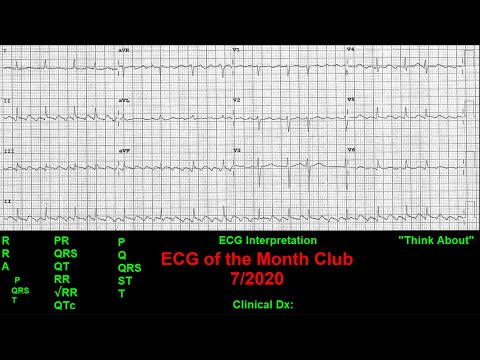
Practice Electrocardiograms Part 1 of 3 (AEI-W1)
During this chapter, we will practice reading electrocardiograms using the ECG reading format outlined in the “Guidelines For Electrocardiogram Interpretation” chapter. I will take you step-by-step through the process and interpret several electrocardiograms.
(ECG #1-3)
Practice Electrocardiograms Part 2 of 3 (AEI-W1)
During this chapter, we will practice reading electrocardiograms using the ECG reading format outlined in the “Guidelines For Electrocardiogram Interpretation” chapter. I will take you step-by-step through the process and interpret several electrocardiograms.
(ECG #4-7)
Practice Electrocardiograms Part 3 of 3 (AEI-W1)
During this chapter, we will practice reading electrocardiograms using the ECG reading format outlined in the “Guidelines For Electrocardiogram Interpretation” chapter. I will take you step-by-step through the process and interpret several electrocardiograms.
(ECG #8-10)
QUIZ - Electrocardiogram (ECG) Basics (AEI-W1)
QUIZ - Lead Placement (AEI-W1)
QUIZ - Axis Determination (AEI-W1)
QUIZ - Atrial abnormalities (AEI-W1)
QUIZ - Ventricular Hypertrophy (AEI-W1)
QUIZ - Bundle Branch Blocks and Intraventricular Conduction Delays (AEI-W1)
QUIZ - Fascicular Blocks (AEI-W1)
QUIZ - Ischemia, Injury, Infarction Part 1 (General findings) (AEI-W1)
QUIZ - Ischemia, Injury, Infarction Part 2 (Pathophysiology) (AEI-W1)
QUIZ - ST and T wave Changes (AEI-W1)
QUIZ - QT Interval (AEI-W1)
QUIZ - Pericarditis (AEI-W1)
QUIZ - Central Nervous System Effects on the ECG (AEI-W1)
QUIZ - Dextrocardia (AEI-W1)
QUIZ - Electrolyte Abnormalities (AEI-W1)
QUIZ - Drug Effects (AEI-W1)
QUIZ - Hypothermia (AEI-W1)
QUIZ - Preexcitation and Wolff Parkinson White [WPW] (AEI-W1)
QUIZ - Lead Reversal (AEI-W1)
QUIZ - Transition (AEI-W1)
QUIZ - Poor R Wave Progression (PRWP) (AEI-W1)
QUIZ - Miscellaneous ECG Findings 1 (AEI-W1)
QUIZ - Miscellaneous ECG Findings 2 (AEI-W1)