All BASIC Course Bundle

Basic Arrhythmia/ECG Interpretation + 40 Practice ECGs + ACLS Rhythm Review + ECG Heart Rhythm Review. **** Bonus - Point of Care Echo

ratings icon 5.0 (3 Reviews)
ratings icon 92+ Videos
ratings icon 2 Exams
ratings icon 63 Quizzes
ratings icon 8+ Hours

ACCESS to ALL BASIC Arrhythmia/ECG Interpretation Courses + All 40 Practice ECGs + ACLS Rhythm Review + ECG Heart Rhythm Review. Bonus - Point of Care Echo Course. You get simplified, and basic ECG and arrhythmia training plus a simplified step-by-step approach to reading ECGs from start to finish. Additionally, you get access to all Practice ECGs with full explanations. All Practice ECGs are read in the same step-by-step approach taught in Advanced ECG Interpretation. As a bonus, you can start learning Point of Care Echocardiography, which is becoming standard in medical practice.

More About This Course

Hide Description

This is the best bundle for beginners learning to read ECG and Arrhythmias. You will walk away with the basic skills for interpreting and recognizing most normal and abnormal heart rhythms, most common ECG abnormalities, and be able to practice on real-life ECG from the ER and clinics. This bundle also prepares you for the rhythms you will see on the ACLS test. As a bonus you will learn the basic of cardiac point of care echocardiography.

Courses Included:

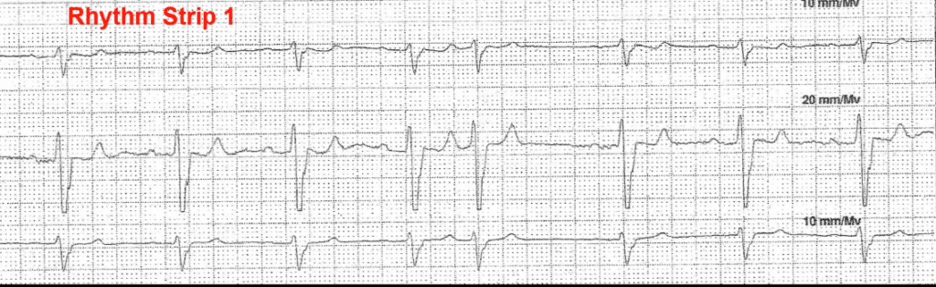
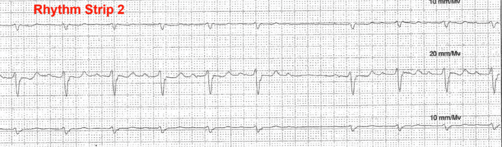
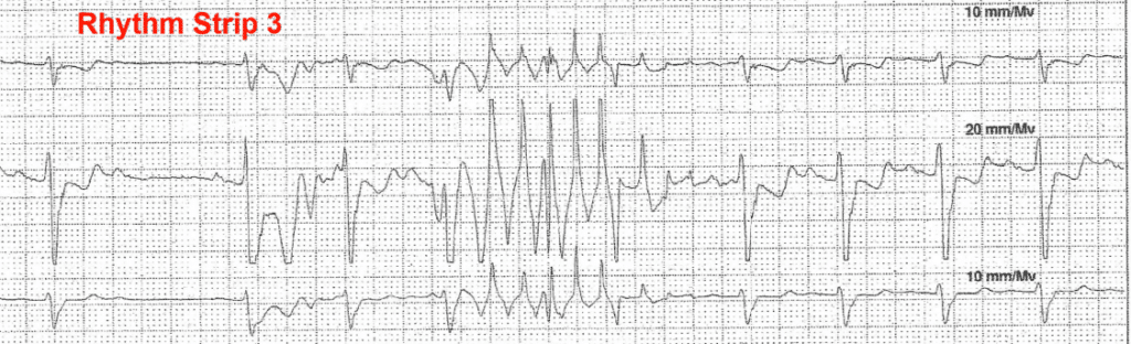
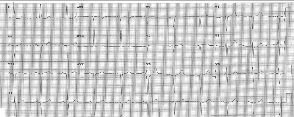
  • Basic Arrhythmia Interpretation – Learn to recognize normal and abnormal heart rhythms with this short, simplified arrhythmia course.
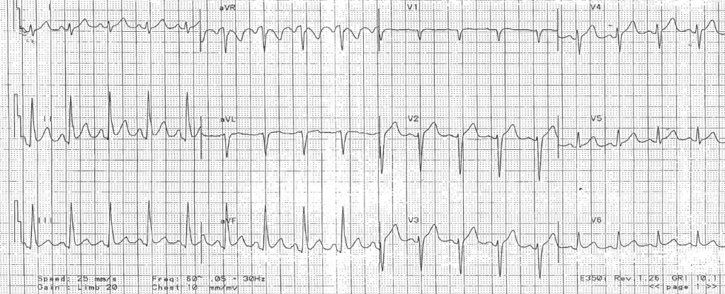
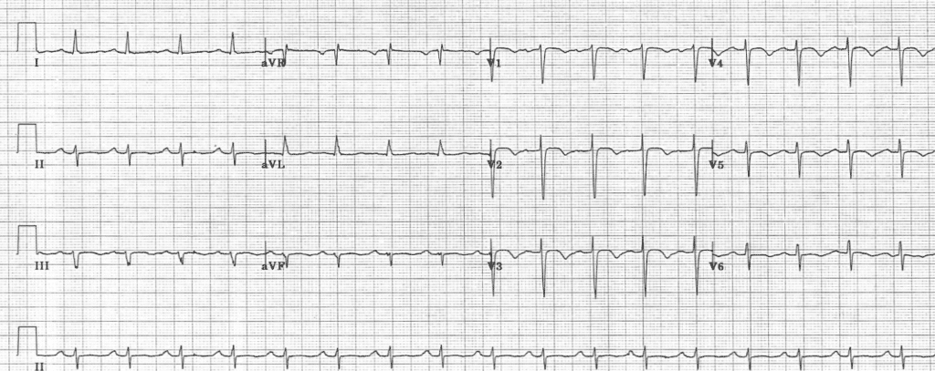
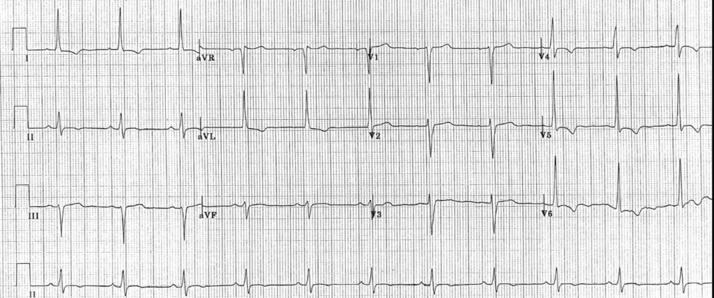
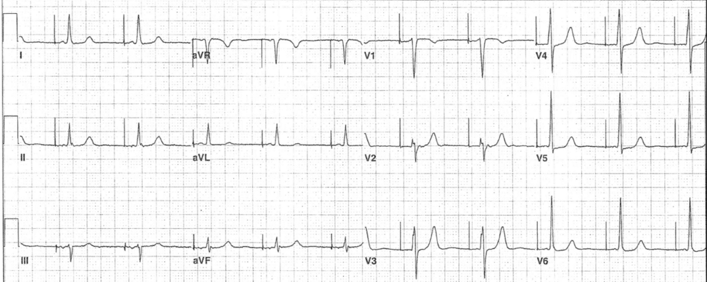
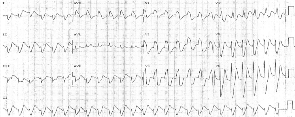
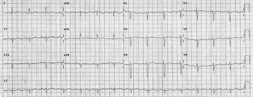
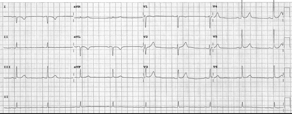
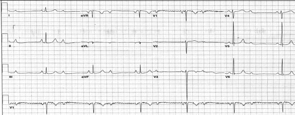
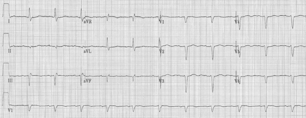
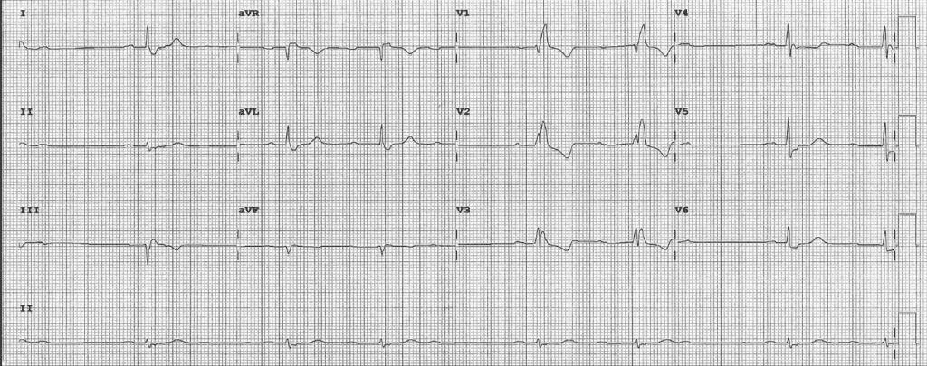
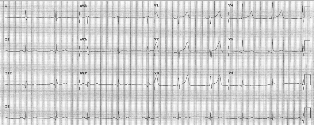
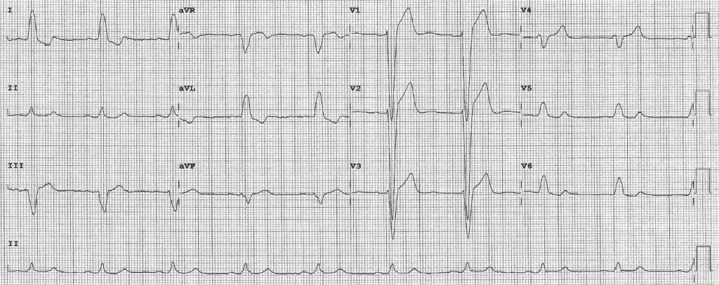
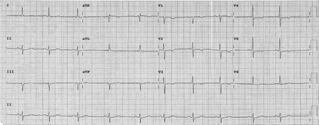
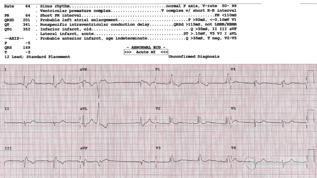
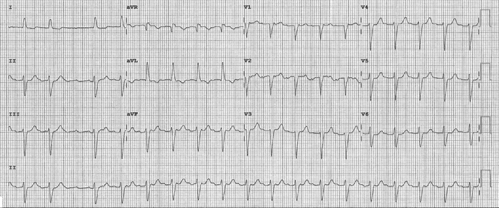
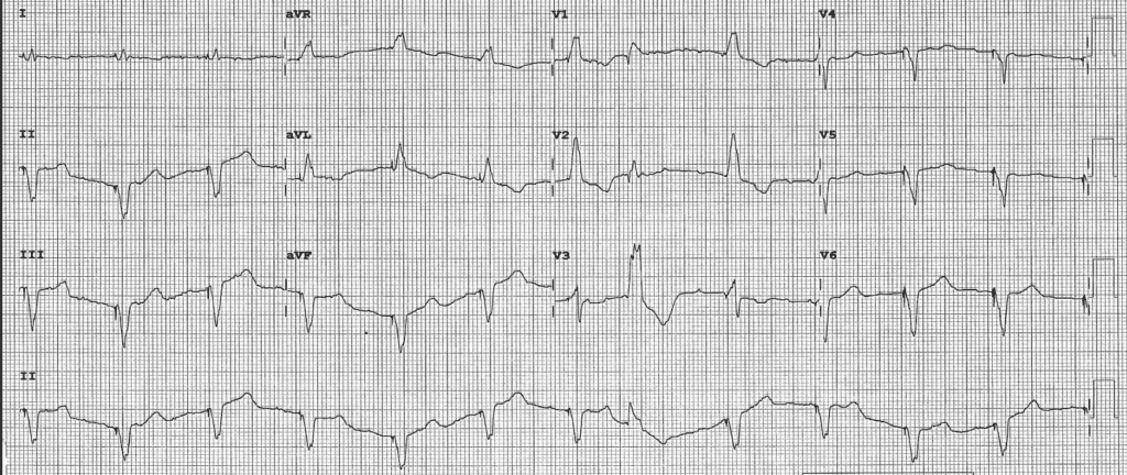
  • Basic ECG Interpretation – Learn the basic normal and abnormal ECG findings, ECG criteria, and a simplified step-by-step approach to reading ECGs.
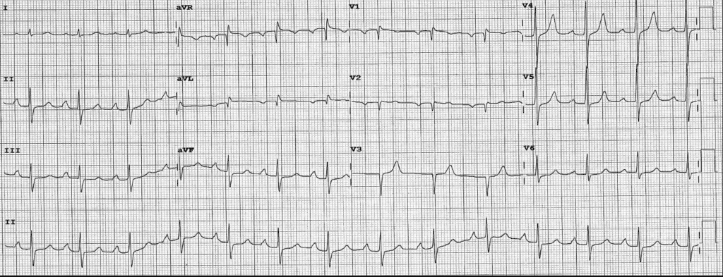
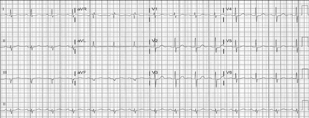
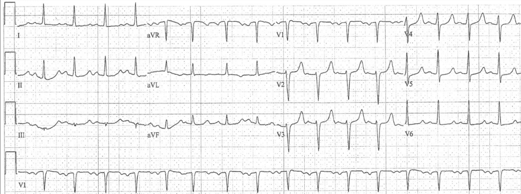
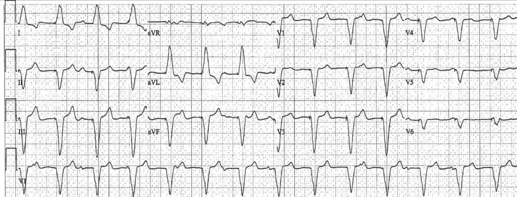
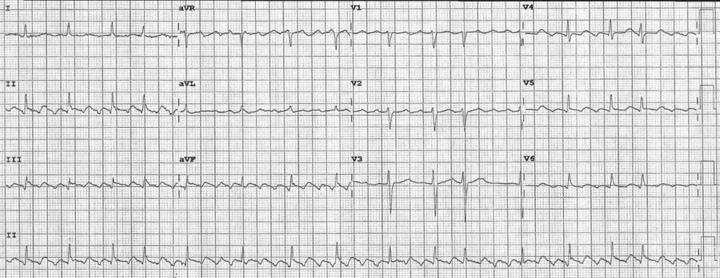
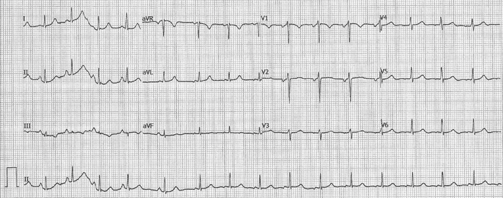
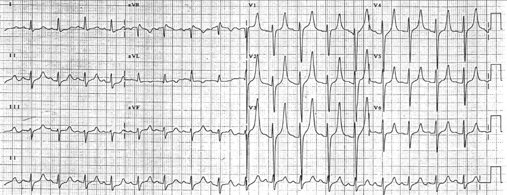
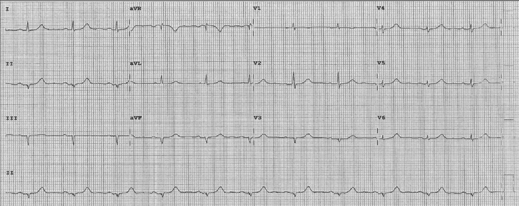
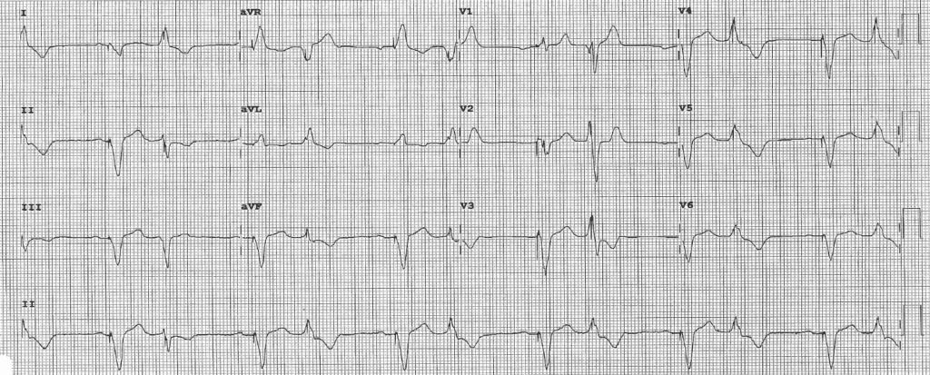
  • Practice ECGs Course – Plenty of practice ECGs that you read yourself, listen to the expert interpretation, and compare your findings. Each ECG is read using the same step-by-step approach taught in Advanced ECG Interpretation.
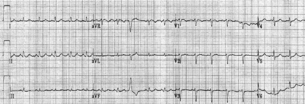
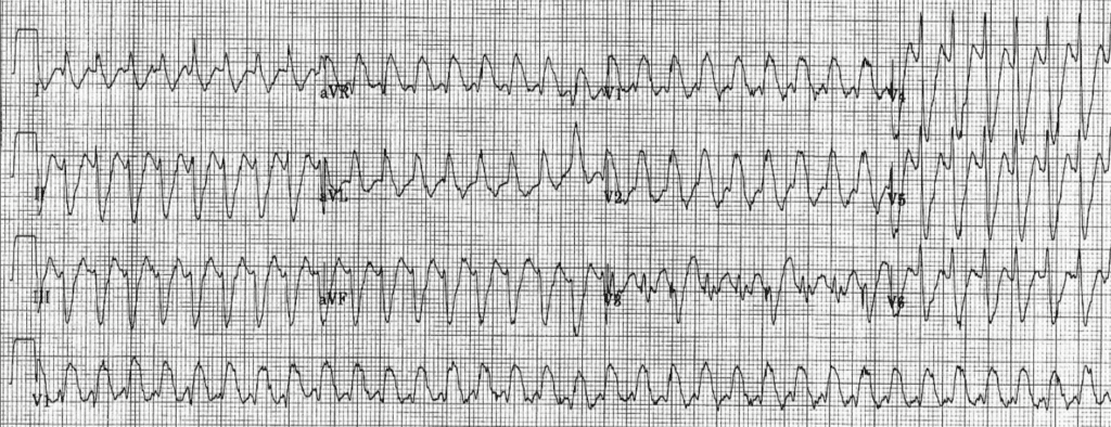
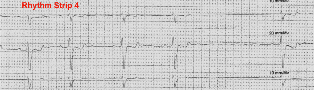
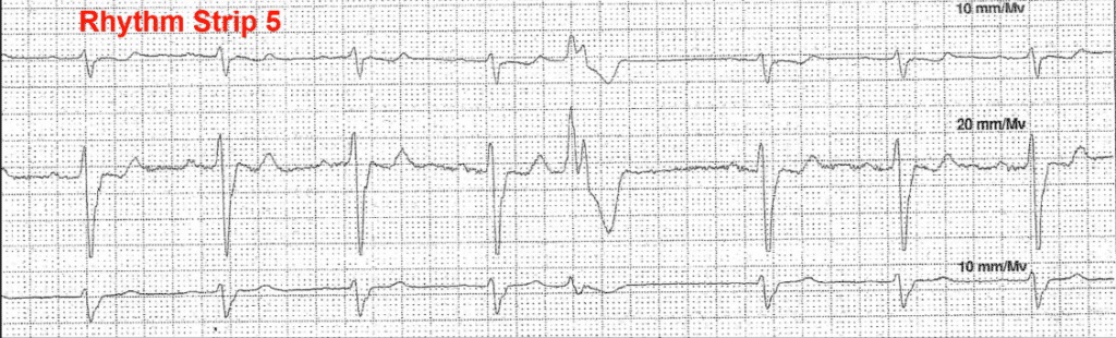
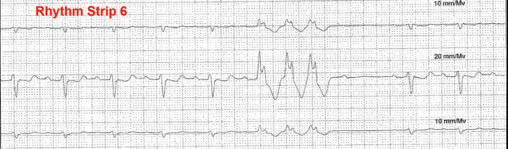
  • ACLS Rhythm Course – All ACLS rhythms are covered in this interactive video. Watch a rhythm strip, identify the rhythm, and then watch and listen to the detailed explanation.
  • ECG Heart Rhythm Review Course – Learn 20 of the most common normal and abnormal heart rhythms in minutes. Just click on the rhythm you want to learn and a video explanation will play.
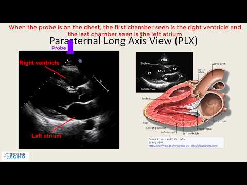
  • Point of Care Echo – Learn cardiac anatomy and physiology through echocardiography. You will learn about the echo probe, the different echo views, the ventricles, the ejection fraction, and the valves. Also included are color flow Doppler, pericardial effusions and tamponade, pleural effusions, mass, vegetations, segmental wall abnormalities, the inferior vena cava, and the aorta.

See each individual course for full details

If you are also interested in learning the finer details and pathophysiology of ECGs and Arrhythmias, please see the All-Access Membership.

Learn More

What's Covered In This Course?

  • Basic ECG/Arrhythmia Criteria
  • A Simple Step-by-Step Approach to Reading ECGs
  • Practice ECGs with detailed explanations of the findings
  • Learn/Review all of the ACLS heart rhythms
  • Certificate of Completion for each course (not including ACLS and ECG Heart Rhythm)
  • 24/7 Access from any device
  • Ability to re-watch the videos over and over

Who This Course Is For

Best for beginners who want it all. Plus enjoy an introduction to echocardiography

Best for:

  • Beginning Medical Student
  • Medical Interns
  • Nurse Practitioners
  • Physician Assistants
  • Nurses
  • EMTs
  • Paramedics
  • Graduate and Undergraduate Students Interested in Medicine

Sample Course Videos

online ecg interpretation course samples
Basic ECG Interpretation - Myocardial ischemia and infarction Watch Preview
online ecg interpretation course samples
Ventricular Rhythms Sample Video Watch Preview
online ecg interpretation course samples
Atrioventricular (AV) Node Blocks Sample Video Watch Preview
online ecg interpretation course samples
Sample Practice ECG Watch Preview
online ecg interpretation course samples
Basic Echo Views - Preview Watch Preview
online ecg interpretation course samples
Free ACLS Rhythm Course Sample Video Watch Preview

Course Syllabi

Basic Arrhythmia Interpretation - Basic Anatomy and Physiology For ECGs (W1)
This chapter reviews the important, basic cardiac anatomy and physiology needed to read electrocardiograms and rhythm strips. This includes the cardiac chambers, valves, electrical system, and circulatory system.
Basic Arrhythmia Interpretation - Action Potential (W1)
This chapter reviews the cardiac muscle action potential.
Basic Arrhythmia Interpretation - How the ECG Machine Sees Electrical Impulses (W1)
This chapter reviews how the ECG machine sees electrical impulses and translates them into the waves seen on the ECG.
Basic Arrhythmia Interpretation - Propagation of Impulses Through the Heart (W1)
This chapter reviews how the cardiac impulse propagates through the heart. Impulses typically start in the sinoatrial (SA) node, go through the atria, the atrioventricular (AV) node, and into the ventricles.
Basic Arrhythmia Interpretation - ECG Calibrations (W1)
This video shows you the ECG calibrations.
Basic Arrhythmia Interpretation - The ECG Waves (W1)
This chapter reviews electrical waveforms. Topics include P-waves, QRS complexes, and T-waves. It reviews Q-waves, R-waves, S-waves, R prime (R'), R double prime (R''), S prime (S'), and S double prime (S'').
Basic Arrhythmia Interpretation - ECG Intervals (W1)
This chapter reviews the intervals of the electrical waveforms. Topics include the PR interval, QRS interval, and QT interval.
Basic Arrhythmia Interpretation - Telemetry Monitoring (W1)
This chapter reviews telemetry monitors and basic telemetry setups.
Basic Arrhythmia Interpretation - Naming Rhythms (W1)
This chapter reviews how to properly name rhythms. It emphasizes that rhythms are named by their location of origin and something about the heart rate.
Basic Arrhythmia Interpretation - Determining the Heart Rate (W1)
This chapter shows several ways to determine the heart rate on an ECG or telemetry strip.
Basic Arrhythmia Interpretation - Sinus Rhythms (W1)
This chapter reviews sinus rhythms (rhythms that originate from the sinoatrial (SA) node). These include normal sinus rhythm, sinus bradycardia, and sinus tachycardia.
Basic Arrhythmia Interpretation - Junctional Rhythms (W1)
This chapter reviews junctional rhythms (rhythms that originate from the atrioventricular (AV) node). These include idiopathic junctional rhythm, junctional bradycardia, accelerated junctional rhythm, and junctional tachycardia.
Basic Arrhythmia Interpretation - Ventricular Rhythms (W1)
This chapter reviews ventricular rhythms (rhythms that originate from the ventricles). These include premature ventricular complexes (PVC), ventricular couplets, monomorphic ventricular tachycardia, polymorphic ventricular tachycardia, torsades de pointes (TdP), and ventricular fibrillation.
Basic Arrhythmia Interpretation - Atrial Rhythms (W1)
This chapter reviews atrial rhythms (rhythms that originate from the atria). These include premature atrial complexes (PAC), ectopic atrial rhythms, atrial bradycardia, atrial tachycardia, atrial flutter, atrial fibrillation, and multifocal atrial tachycardia.
Basic Arrhythmia Interpretation - Supraventricular Tachycardia [SVT] (W1)
This chapter reviews supraventricular tachycardia. specifically, it covers a fast rhythm that originates from the atrioventricular (AV) node.
Basic Arrhythmia Interpretation - Asystole and Agonal Rhythms (W1)
This chapter reviews asystole (no heart rhythm) and agonal rhythms (slow variable ventricular rhythms of a dying heart).
Basic Arrhythmia Interpretation - Pacemaker Rhythms (W1)
This chapter reviews pacemaker rhythms (rhythms that originate from a pacemaker). These include ventricular pacemaker rhythms and atrioventricular pacemaker rhythms.
Basic Arrhythmia Interpretation - ECG Artifacts (W1)
This video goes over possible artifacts seen on the ECG and rhythm strips.
Basic Arrhythmia Interpretation - Atrioventricular (AV) Blocks (W1)
This lesson goes over atrioventricular (AV) blocks. It covers first-degree AV blocks, second-degree AV blocks (Mobitz 1 [Wenckebach] and Mobitz 2), third-degree (complete) AV blocks, and 2:1 AV blocks
Basic Arrhythmia Interpretation - Serious vs. Non-serious Rhythms (W1)
This chapter briefly reviews which heart rhythms are serious and may need immediate medical attention, and which rhythms are overall benign.
Basic ECG Interpretation - The 12-Lead ECG (W1)
This lesson tells about ECGs and how the ECG machine translates the electrical activity of the heart into an ECG graph.
Basic ECG Interpretation - Correct ECG Lead Placement (W1)
This video shows you the proper position to place the electrodes on a patient to obtain an ECG.
Basic ECG Interpretation - The QRS Axis (W1)
This lesson teaches how to calculate the QRS axis and determine if it is normal. It reviews conditions that cause left axis deviation, right axis deviation, and superior axis.
Basic ECG Interpretation - Basic Cardiac Rhythms (W1)
Although this material is covered in the arrhythmia section of this course, this video gives a great summary of normal and abnormal cardiac rhythms in a single, short video.
Basic ECG Interpretation - Left and Right Atrial Abnormality (W1)
This video teaches a quick and easy way to identify left and right atrial abnormality.
Basic ECG Interpretation - Left and Right Ventricular Hypertrophy (W1)
This chapter teaches how to identify left and right ventricular hypertrophy on the ECG. It also describes secondary ST and T wave changes that might accompany these conditions.
Basic ECG Interpretation - Bundle Branch Blocks, Conduction Delays, and Fascicular Blocks (W1)
This video teaches how to identify left and right bundle branch blocks, non-specific intraventricular conduction delays, and left anterior- and left posterior-fascicular blocks.
Basic ECG Interpretation - Myocardial Ischemia and Infarction (W1)
This important lesson shows how to identify myocardial ischemia and myocardial infarctions. It shows how to diagnose an acute myocardial infarction and then goes through the evolutionary ST and T changes that occur over time. You'll learn to recognize acute, recent, age-undetermined, and old myocardial infarctions on the ECG.
Basic ECG Interpretation - ST and T Wave Changes (W1)
This lesson teaches the differences between primary ST and T wave changes, as seen in ischemia, from secondary ST and T wave changes, as seen in conditions like left ventricular hypertrophy.
Basic ECG Interpretation - Low QRS Voltage (W1)
This video teaches you to recognize low QRS voltage and then gives you a brief differential diagnosis of urgent/emergent conditions that cause low QRS voltage. This is important to recognize since some of the causes of low voltage are life-threatening and need immediate attention.
Basic ECG Interpretation - ECG Changes From Electrolyte Abnormalities (W1)
This video teaches about ECG changes from hyperkalemia, hypokalemia, hypercalcemia, and hypocalcemia.
Basic ECG Interpretation - Drug Effects on the ECG and Prolonged QT Interval (W1)
This video goes over the effects of digoxin on the ECG and reviews several medication classes that prolong the QT interval.
Basic ECG Interpretation - ECG Changes from Wolff Parkinson White (WPW) (W1)
This section teaches how to identify the ECG changes from preexcitation and Wolff Parkinson White (WPW) Syndrome.
Basic ECG Interpretation - Guidelines to Reading ECGs (W1)
This chapter shows you a step-by-step approach to reading ECGs and takes you through an example.
Definitions of Point of Care Echocardiography and Objectives (W1)
This chapter defines point of care echocardiography and the objectives of this video course
Basic Cardiac Anatomy (W1)
This chapter reviews cardiac anatomy. It shows the heart in the chest and the position of all four (4) cardiac chambers in relationship to the whole heart and chest cavity.
Understanding the Probe (Transducer) and Probe Movements (W1)
This chapter takes a close look at the Probe/Transducer: Topics include probe anatomy (head, tail, and notch); and probe movements (rocking, tilting, rotating, sliding, and translation).
Basic Echocardiographic Views (W1)
This chapter reviews the different echocardiographic views. Topics include:  Parasternal long axis; right ventricular inflow tract; right ventricular outflow tract; parasternal short axis at the levels of the mid ventricle, mitral valve, and aortic valve; apical four-chamber; apical five-chamber; apical two-chamber; apical three-chamber; subcostal; inferior vena cava; hepatic vein; and suprasternal.
Cardiac Chamber Sizes and Wall Thickness (W1)
This chapter review general cardiac chamber sizes and left and right ventricular wall thickness. Topics include Normal and abnormal left ventricular and right ventricular size and wall thickness; normal and abnormal atrial sizes; and aortic root size.
Identifying the Different Left Ventricular Walls (W1)
This chapter details the different segments and walls of the left ventricle. Topics include Anterior wall; anterior and inferior septum; inferior wall; and anterior and inferior lateral wall.
Left Ventricular Function and Ejection Fraction (W1)
This chapter reviews left ventricular function and the ejection fraction. Topics include Contractility and wall motion; normal systolic function; systolic dysfunction; hyperdynamic function; ejection fraction; and segmental wall motion abnormalities.
Right Heart Function and Abnormalities (W1)
This chapter reviews right ventricular size and function. Topics include Normal right ventricular function; right ventricular dysfunction; right ventricular pressure and volume overload; and McConnell’s sign.
Inferior Vena Cava (W1)
This chapter examines the inferior vena cava. Topics include Normal and abnormal inferior vena cava size and inspiratory collapse; and estimating the right atrial pressure.
Color Flow Doppler (W1)
This chapter discusses and shows examples of color flow Doppler.
Valvular Abnormalities (W1)
This chapter details and shows examples of various valvular abnormalities. Topics include Mitral stenosis; mitral regurgitation; aortic stenosis; aortic regurgitation; and tricuspid regurgitation.
Masses and Vegetations (W1)
This chapter shows how to identify various cardiac masses and vegetations.
Pericardial Effusions, Cardiac Tamponade and Pleural Effusions (W1)
This chapter discusses and shows examples of pericardial effusions, cardiac tamponade, and pleural effusions.
Aortic Aneurysms and Aortic Dissections (W1)
This chapter defines and shows examples of aortic aneurysms and aortic dissections.
Approach to Reading Point of Care Echocardiograms (W1)
This chapter explains my approach to reading echocardiograms accurately and efficiently.
Practice ECG 1 (W1)

Read this ECG yourself and then listen to the detailed explanation of the ECG findings.


[caption id="attachment_13744" align="alignnone" width="1100"] Practice ECG 1 Practice ECG 1[/caption]
Practice ECG 2 (W1)

Read this ECG yourself and then listen to the detailed explanation of the ECG findings.


[caption id="attachment_13745" align="alignnone" width="1100"] Practice ECG 2 Practice ECG 2[/caption]
Practice ECG 3 (W1)

Read this ECG yourself and then listen to the detailed explanation of the ECG findings.


[caption id="attachment_13746" align="alignnone" width="1100"] Practice ECG 3 Practice ECG 3[/caption]
Practice ECG 4 (W1)

Read this ECG yourself and then listen to the detailed explanation of the ECG findings.


[caption id="attachment_13747" align="alignnone" width="1100"] Practice ECG 4 Practice ECG 4[/caption]
Practice ECG 5 (W1)

Read this rhythm strip yourself and then listen to the detailed explanation of the findings.


On rhythm strips you only read the rhythm and the intervals. Do not try to do a full ECG interpretation as there is not enough data.


[caption id="attachment_13748" align="alignnone" width="1100"] Practice ECG 5 Practice ECG 5[/caption]
Practice ECG 6 (W1)


Read this ECG yourself and then listen to the detailed explanation of the ECG findings.


[caption id="attachment_13749" align="alignnone" width="1100"] Practice ECG 6 Practice ECG 6[/caption]
Practice ECG 7 (W1)


This is an ECG of a 28-year-old man in the Emergency Department with possible food poisoning. Read it yourself and then listen to the detailed explanation of the ECG findings.


[caption id="attachment_13750" align="alignnone" width="1100"] Practice ECG 7 Practice ECG 7[/caption]
Practice ECG 8 (W1)

Read this ECG yourself and then listen to the detailed explanation of the ECG findings.


[caption id="attachment_13751" align="alignnone" width="1100"] Practice ECG 8 Practice ECG 8[/caption]
Practice ECG 9 (W1)

This is an ECG of a 60-year-old woman. Read it yourself and then listen to the detailed explanation of the ECG findings.


[caption id="attachment_13752" align="alignnone" width="1100"] Practice ECG 9 Practice ECG 9[/caption]
Practice ECG 10 (W1)

Read this ECG yourself and then listen to the detailed explanation of the ECG findings.


[caption id="attachment_13753" align="alignnone" width="1100"] Practice ECG 10 Practice ECG 10[/caption]
Practice ECG 11 (W1)

Here is an interesting ECG. Read it yourself and then listen to the detailed explanation of the ECG findings. In the end, clinical patient information is shared.


[caption id="attachment_13754" align="alignnone" width="1100"] Practice ECG 11 Practice ECG 11[/caption]
Practice ECG 12 (W1)

Here are six rhythm strips from a 48-hour Holter monitor. These strips were from a 76-year-old man being assessed for tachycardia. Each rhythm strip has a unique finding. Read each one and then listen to the detailed explanation. Different reading methods are used to assess the strips.

Remember to click on "Mark Complete" at the bottom of this page when you completed the lesson


[caption id="attachment_13755" align="alignnone" width="1100"] Practice ECG 12 - Rhythm Strip 1 Practice ECG 12 - Rhythm Strip 1[/caption]
[caption id="attachment_13756" align="alignnone" width="1100"] Practice ECG 12 - Rhythm Strip 2 Practice ECG 12 - Rhythm Strip 2[/caption]
[caption id="attachment_13757" align="alignnone" width="1100"] Practice ECG 12 - Rhythm Strip 3 Practice ECG 12 - Rhythm Strip 3[/caption]
[caption id="attachment_13758" align="alignnone" width="1100"] Practice ECG 12 - Rhythm Strip 4 Practice ECG 12 - Rhythm Strip 4[/caption]
[caption id="attachment_13759" align="alignnone" width="1100"] Practice ECG 12 - Rhythm Strip 5 Practice ECG 12 - Rhythm Strip 5[/caption]
[caption id="attachment_13760" align="alignnone" width="1100"] Practice ECG 12 - Rhythm Strip 6 Practice ECG 12 - Rhythm Strip 6[/caption]
Practice ECG 13 (W1)

This ECG is from a 60-year-old man. Read it yourself and then listen to the detailed explanation of the ECG findings.


[caption id="attachment_13761" align="alignnone" width="1100"] Practice ECG 13 Practice ECG 13[/caption]
Practice ECG 14 (W1)

Read this ECG yourself and then listen to the detailed explanation of the ECG findings.


[caption id="attachment_13762" align="alignnone" width="1100"] Practice ECG 14 Practice ECG 14[/caption]
Practice ECG 15 (W1)

Read this ECG yourself and then listen to the detailed explanation of the ECG findings. In the end, clinical patient information is shared.


[caption id="attachment_13763" align="alignnone" width="1024"] Practice ECG 15 Practice ECG 15[/caption]
Practice ECG 16 (W1)

Read this ECG yourself and then listen to the detailed explanation of the ECG findings. In the end, clinical patient information is shared.


[caption id="attachment_13764" align="alignnone" width="1100"] Practice ECG 16 Practice ECG 16[/caption]
Practice ECG 17 (W1)

Read this ECG yourself and then listen to the detailed explanation of the ECG findings.


[caption id="attachment_13765" align="alignnone" width="1100"] Practice ECG 17 Practice ECG 17[/caption]
Practice ECG 18 (W1)

This ECG is from a 40-year-old woman. Read it yourself and then listen to the detailed explanation of the ECG findings.


[caption id="attachment_13766" align="alignnone" width="1100"] Practice ECG 18 Practice ECG 18[/caption]
Practice ECG 19 (W1)


Read this ECG yourself and then listen to the detailed explanation of the ECG findings.


[caption id="attachment_13767" align="alignnone" width="1100"] Practice ECG 19 Practice ECG 19[/caption]
Practice ECG 20 (W1)

Read this ECG yourself and then listen to the detailed explanation of the ECG findings.


[caption id="attachment_13768" align="alignnone" width="1100"] Practice ECG 20 Practice ECG 20[/caption]
Practice ECG 21 (W1)

Read this ECG yourself and then listen to the detailed explanation of the ECG findings.


[caption id="attachment_13769" align="alignnone" width="1100"] Practice ECG 21 Practice ECG 21[/caption]
Practice ECG 22 (W1)

Read this ECG yourself and then listen to the detailed explanation of the ECG findings.


[caption id="attachment_13770" align="alignnone" width="1100"] Practice ECG 22 Practice ECG 22[/caption]
Practice ECG 23 (W1)

Check out this ECG. There is a lot here. Be sure to follow the Executive Electrocardiogram Education ECG Reading Approach to be sure you do not miss anything. Pay close attention to the P-waves and the rhythm. Have fun and then listen to the detailed explanation of the ECG findings.


[caption id="attachment_13771" align="alignnone" width="1100"] Practice ECG 23 Practice ECG 23[/caption]
Practice ECG 24 (W1)

This ECG is from a 78-year-old man with substernal chest pressure. Read it yourself and then listen to the detailed explanation of the ECG findings.


[caption id="attachment_13772" align="alignnone" width="1100"] Practice ECG 24 Practice ECG 24[/caption]
Practice ECG 25 (W1)

This ECG is from a 53-year-old man who presented to the emergency room with palpitations. Read it yourself and then listen to the detailed explanation of the ECG findings.


[caption id="attachment_13773" align="alignnone" width="1100"] Practice ECG 25 Practice ECG 25[/caption]
Practice ECG 26 (W1)

This ECG is from a 77-year-old man who presented to the emergency room with a cough. Read it yourself and then listen to the detailed explanation of the ECG findings.


[caption id="attachment_13774" align="alignnone" width="1100"] Practice ECG 26 Practice ECG 26[/caption]
Practice ECG 27 (W1)

This ECG is from a 26-year-old woman with a history of palpitations and near-syncope. Pay close attention to the rhythm. Read it yourself and then listen to the detailed explanation of the ECG findings.


[caption id="attachment_13775" align="alignnone" width="1100"] Practice ECG 27 Practice ECG 27[/caption]
Practice ECG 28 (W1)

There was no clinical information at the time the ECG was received. Read it yourself and then listen to the detailed explanation of the ECG findings.


[caption id="attachment_13776" align="alignnone" width="1100"] Practice ECG 28 Practice ECG 28[/caption]
Practice ECG 29 (W1)

Read this ECG yourself and then listen to the detailed explanation of the ECG findings.


[caption id="attachment_13777" align="alignnone" width="1100"] Practice ECG 29 Practice ECG 29[/caption]
Practice ECG 30 (W1)

Read this ECG yourself and then listen to the detailed explanation of the ECG findings.


[caption id="attachment_13778" align="alignnone" width="1100"] Practice ECG 30 Practice ECG 30[/caption]
Practice ECG 31 (W1)

This ECG is from a 70-year old man. There is no clinical history. Read it yourself and then listen to the detailed explanation of the ECG findings.


[caption id="attachment_13779" align="alignnone" width="1100"] Practice ECG 31 Practice ECG 31[/caption]
Practice ECG 32 (W1)

Please read this ECG from an asymptomatic 69-year-old man, seen in the cardiology office for a routine check-up. When you are done listen to the detailed explanation of the ECG findings.


[caption id="attachment_13780" align="alignnone" width="1100"] Practice ECG 32 Practice ECG 32[/caption]
Practice ECG 33 (W1)

This ECG is from an 85-year old woman seen during a routine cardiology office visit. She is asymptomatic. Read it yourself and then listen to the detailed explanation of the ECG findings.


[caption id="attachment_13781" align="alignnone" width="1100"] Practice ECG 33 Practice ECG 33[/caption]
Practice ECG 34 (W1)

This ECG is from a 71-year old man who is asymptomatic. Read this ECG and then listen to the detailed explanation of the ECG findings.


[caption id="attachment_13782" align="alignnone" width="1100"] Practice ECG 34 Practice ECG 34[/caption]
Practice ECG 35 (W1)

This ECG is from a 77-year-old woman. Read it yourself and then listen to the detailed explanation of the ECG findings.


[caption id="attachment_13783" align="alignnone" width="1100"] Practice ECG 35 Practice ECG 35[/caption]
Practice ECG 36 (W1)

With this ECG, we will take a completely different approach to the interpretation. I want you to look at the diagnoses made by the computer, and see if you agree or disagree. You need to defend your positions. After making your decisions, listen to the detailed explanations. You may want to read the ECG from beginning to end, as usual, and make your final diagnoses. This ECG is from an asymptomatic 84-year-old woman seen in the office.


[caption id="attachment_13739" align="alignnone" width="1100"] Practice ECG 36 Practice ECG 36[/caption]
Practice ECG 37 (W1)

Read it yourself and then listen to the detailed explanation of the ECG findings.


[caption id="attachment_13740" align="alignnone" width="1100"] Practice ECG 37 Practice ECG 37[/caption]
Practice ECG 38 (W1)

This ECG is from a 89-year old man during a routine office visit. Read it yourself and then listen to the detailed explanation of the ECG findings.


[caption id="attachment_13741" align="alignnone" width="1100"] Practice ECG 38 Practice ECG 38[/caption]
Practice ECG 39 (W1)

This ECG is from a 70-year-old man seen in the office. Read it yourself and then listen to the detailed explanation of the ECG findings.


[caption id="attachment_13742" align="alignnone" width="1100"] Practice ECG 39 Practice ECG 39[/caption]
Practice ECG 40 (W1)

This ECG is from a 75-year-old woman seen in a cardiology office setting. Read it yourself and then listen to the detailed explanation of the ECG findings.


[caption id="attachment_13743" align="alignnone" width="1100"] Practice ECG 40 Practice ECG 40[/caption]

Feedback From Our Subscribers

Alan Ghaly, DO, FACC Verified Subscriber
five star rating
Informative and easy to understand!

I had the pleasure of being lectured about electrocardiogram interpretation by Dr. Siegal during my cardiology training. These videos are just like his lectures - informative and easy to understand in a field that can be intimidating. I wish I had these videos during my medical school and training years.

V. Richardson Verified Subscriber
five star rating
Great visual examples

The videos are great for people who are visual and audio learners. They go at a pace that is easy to understand and there are good visual examples.

Michael Friedman, DO, Assistant Professor, Rowan University School of Osteopathic Medicine Verified Subscriber
five star rating
Dr. Siegal shared his knowledge

"Dr. Siegal is a doctor's doctor. He has taken his clinical knowledge and experience and thankfully shared it with us in an informative and well written video."