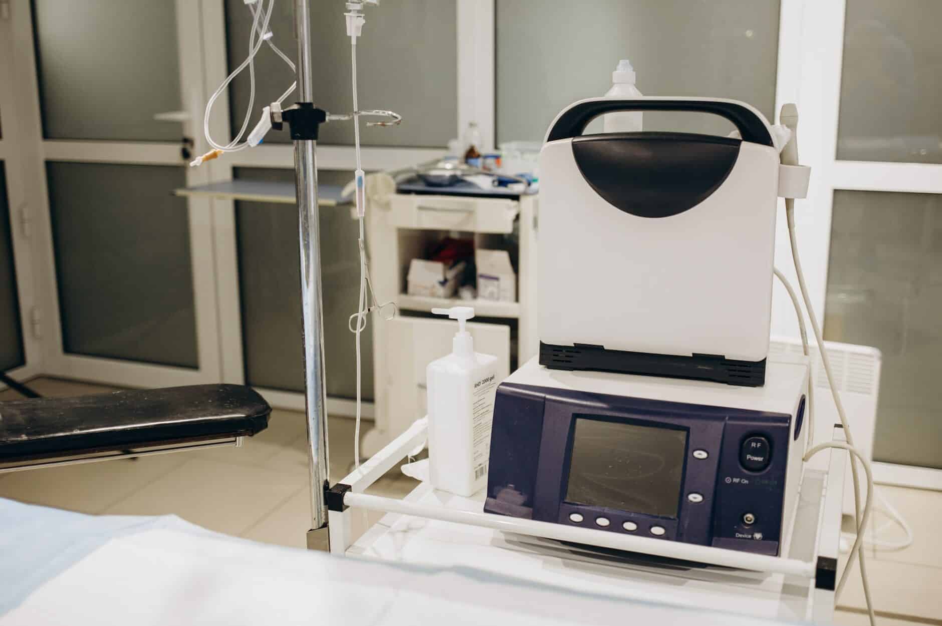
- Echocardiography’s Role In Treating Heart Disease
- Basics Of Echocardiographic Images
- Echocardiographic Techniques To Improve Image Quality
- Advanced Techniques For Echocardiographic Imaging
- Common Challenges In Echocardiographic Imaging
- Quality Control And Quality Assurance
- Start Improving Your Echocardiography Skills Today
Every cardiologist knows that obtaining high-quality echocardiographic images is critical in establishing an accurate diagnosis and creating an effective treatment plan for patients.
However, in many ways, echocardiographic imaging is as much an art as it is a science, so it’s important to understand the different factors that can impact image quality and to have a toolbelt of techniques to account for them.
Keep reading for a deeper dive into standard views, probe positions, advanced technologies, common imaging challenges, and more.
Echocardiography’s Role In Treating Heart Disease
Echocardiography is a noninvasive imaging technique that uses ultrasound waves to produce high-quality images of the heart and surrounding structures, providing physicians with valuable information about cardiac function and pathology.
Applications For Echocardiography
Echocardiography plays a central role in evaluating cardiac function, valvular abnormalities, wall motion abnormalities, and other cardiovascular pathologies.
Echocardiography’s importance cannot be overstated. Its ability to provide detailed information about the anatomy and physiology of the heart without exposing patients to ionizing radiation or invasive procedures makes it an indispensable tool in any clinician’s arsenal.
This increases patient safety and facilitates effective monitoring and tracking of disease progression.
Benefits Of High-Quality Echocardiographic Images
High-quality echocardiographic images are critical in accurately preventing, diagnosing, and treating cardiovascular diseases. By providing clear and comprehensive views of the structures and functions of the heart, these images enable medical professionals to make informed decisions regarding patient care while minimizing the need for invasive diagnostic procedures.
Additionally, high-quality echocardiographic images also improve patient outcomes by allowing early detection of potential ailments before they become serious or life-threatening.
Cases such as pediatric coronary artery imaging demonstrate the importance of using the correct settings and thoroughly understanding ultrasound physics to achieve optimal results.
Higher-quality images increase diagnostic accuracy and reduce the risk of misdiagnosis, ultimately leading to more effective treatment plans tailored specifically to the needs of individual patients.
Basics Of Echocardiographic Images
Like in any other area of medicine, mastering the fundamentals is a critical first step in improving your technique. In echocardiography, you should understand the standard views, the role that image orientation plays, and the factors that affect image quality in echocardiography.
Standard Views And Image Orientation
Standard views and image orientation are fundamental aspects of echocardiographic imaging that must be mastered to ensure accurate diagnostic results. They provide the framework for a consistent and systematic approach to cardiac examination and allow medical personnel to obtain good-quality images during the exam.
To navigate these standard views, physicians must be familiar with anatomic landmarks such as papillary muscles, great vessels, valves, and ventricular walls.
Familiarity with different probe positions also plays a critical role in obtaining optimal images representative of specific regions of the heart. For example, the parasternal long-axis view shows structures such as the left atrium, mitral valves, and subvalvular apparatus. In contrast, the apical four-chamber view provides a view of all four chambers and the anatomy of the tricuspid valve.
Factors Affecting Image Quality
Several factors can influence image quality, including patient-specific variables such as body habitus, lung disease, and cardiac anatomy.
The imaging settings selected for echocardiography are key to obtaining high-quality images. These settings should be chosen based on factors such as the type of image to be acquired, the desired resolution, and the size and anatomy of the patient.
Imaging parameters should be set to optimize image quality, taking into account variables such as depth, gain, dynamic range, and image persistence.
Additionally, color Doppler imaging can further improve image accuracy. It’s important to set parameters to ensure the best possible image quality and accuracy for the imaging application.
Echocardiographic Techniques To Improve Image Quality
Physicians can utilize several techniques to consistently obtain high-quality images and overcome challenges presented by patient-specific variables.
Some of the most impactful include patient preparation, proper probe placement and angulation, and optimization of imaging settings to obtain high-quality echocardiographic images.
Patient Preparation
Proper patient prep is the first and, possibly, most important step in this process. As you know, image quality depends on more than probe placement alone. Here are a few important steps to focus on.
- Explain to the patient what will happen during the procedure. Make sure to include the purpose, duration, and possible risks.
- Ask the patient about any medical conditions or medications they’re taking that could affect image quality or safety during the exam.
- Place the patient appropriately, lying them flat on their back with their left side slightly up. Ensure that the patient’s clothing doesn’t obstruct the transducer’s path.
- Attach ECG electrodes to monitor heart rhythm and respiratory effort during imaging.
- Provide pillows or cushions for comfort and support as needed.
- Instruct patients not to eat or drink anything for at least 2 hours before the exam, if possible, to avoid bloating in the stomach or intestines, as these could interfere with ultrasound transmission.
Probe Placement And Angulation
After patient preparation, probe placement and angulation are the next most important factor in image quality. Here are a few techniques to remember for this part of the process.
- Identify the proper acoustic window by visualizing the chest wall and locating the intercostal spaces.
- Place the probe perpendicular to the ribs and parallel to the intercostal spaces for optimal imaging.
- It’s best to start with a standard view allowing consistent imaging evaluation.
- Adjust the probe orientation and location to capture different cardiac structures, for example, by adjusting the imaging depth or angulating it slightly toward the right shoulder.
- Remember that even slight changes in probe position can significantly improve or worsen image quality.
Optimization Of Imaging Settings
The next factor to consider is the image settings being used. Proper optimization of imaging settings can help detect pathology that may have previously gone undetected, allowing the physician to intervene early. Here are a few things to keep in mind.
- Adjust Gain to achieve optimal grayscale and contrast values.
- Optimize Time gain compensation (TGC) settings to adjust signal quality as it travels through different tissue depths. Optimize these settings for each patient to reduce image artifacts.
- Adjust depth and focus according to the target structure to ensure clear visualization of all cardiac structures.
- Optimize color Doppler settings for accurate flow detection and ensure minimal ghosting or noise in the image.
- Use harmonic imaging to reduce image artifacts and increase the depth of penetration.
- Use the zoom feature to capture smaller structures more accurately.
Advanced Techniques For Echocardiographic Imaging
Thanks to the advancements in processing power, digital imaging, and medical software, new advanced techniques in echocardiographic imaging have emerged in recent years. Among other benefits, these techniques enable medical professionals to obtain more detailed and accurate cardiac structure and function assessments.
Three-Dimensional Echocardiography
In recent years, three-dimensional echocardiography (3DE) has become an important tool for diagnosing and treating cardiac diseases. Unlike traditional two-dimensional echocardiography, 3DE allows physicians to view the heart from any angle, providing a more detailed and accurate assessment of its structure and function.
This technology has many potential applications, such as transcatheter valve replacement or repair surgery. It can also detect and diagnose medical conditions quicker and more accurately and monitor patients’ progress during and after treatment.
The transesophageal 3D echocardiogram (3D-TEE) can also be used to create patient-specific anatomic models for preoperative planning. By creating a three-dimensional model of the patient’s anatomy, surgeons can visualize the procedure before it’s begun, allowing them to plan for any potential surprises.
Contrast Echocardiography
Contrast echocardiography is a technique widely used in cardiology that helps improve image quality, reader confidence, and reproducibility. By introducing microbubbles into the bloodstream, contrast agents enhance the visualization of cardiac structures and increase the accuracy of diagnostic information.
The American Society of Echocardiography has published a guide to the clinical use of contrast agents, which includes recommendations for their use in various clinical scenarios.
Recent technological advances have also led to new formulations and delivery methods for contrast agents that further improve their safety profile and efficacy. Therefore, physicians need to keep abreast of these advances and incorporate them into their routine practice.
Strain Imaging
Strain imaging is another advanced technique used to assess the function of the heart muscle. It involves measuring the deformation or change in the heart muscle during different phases of the cardiac cycle.
Echocardiograms with strain measurement are becoming more common because they provide physicians with more comprehensive information about a patient’s cardiovascular health.
Other Emerging Technologies
In addition to three-dimensional echocardiography and contrast echocardiography, other new technologies can improve the quality of echocardiographic imaging.
A new technology is speckle-tracking echocardiography (STE). STE is a state-of-the-art technique that analyzes the movement of speckles, natural acoustic markers, in the myocardium. This method allows for myocardial strain quantification and aids in the early detection of cardiac dysfunction.
Common Challenges In Echocardiographic Imaging
While every patient is different, there are a few typical situations that you may run into when obtaining echocardiographic images.
Obese Patients And Limited Acoustic Windows
In obese patients, echocardiographic imaging can be challenging because of poor acoustic windows and attenuation artifacts. Rapid and efficient echocardiographic image acquisition is required to ensure optimal diagnostic sensitivity in these patients.
To overcome these difficulties, there are several techniques that physicians can consider:
- Use low-frequency probes to achieve deeper penetration and enhance anechoic window visualization.
- Optimize the probe pressure on the chest wall to ensure better signal transmission.
- Adjust gain settings on the ultrasound machine to compensate for increased tissue attenuation.
- Consider transesophageal echocardiography as an alternative when transthoracic echocardiography has limited visibility.
- In cases where stress testing is needed, consider a nuclear stress test instead of an echo stress test.
- In obese patients, weight loss prior to cardiac imaging may also be considered to improve image quality.
Patients With Lung Disease
Echocardiographic imaging can be especially tricky for patients with pulmonary disease. However, the scans’ accuracy is critical to understanding the individual risk factors of the patient’s condition. Here are some techniques that can be used:
- Patients with respiratory distress should be positioned at a 45-degree angle with their hands behind their heads. This will help expand the lungs and improve acoustic windows.
- In patients with severe lung disease, transthoracic echocardiography (TTE) may not be possible or may provide inadequate images. Transesophageal echocardiography (TEE) is an alternative that can provide clearer images.
- Adjust TTE settings to achieve maximum penetration and enhancement without compromising image quality.
- In cases where standard TTE methods are inadequate, contrast agents provide a better signal-to-noise ratio.
- Respiratory therapists can help by adjusting ventilator settings during imaging, coordinating oxygen administration, or interpreting ventilator perfusion scans for more accurate diagnoses.
Quality Control And Quality Assurance
Quality control (QC) and quality assurance (QA) are vital to ensure consistent, accurate, and reliable diagnostic results. Implementing effective QC and QA measures can improve patient care, optimize resource usage, and reduce the risk of misdiagnosis or suboptimal management.
Importance Of Quality Control And Assurance
Quality control and assurance are essential components of high-quality echocardiographic imaging. This involves ensuring that equipment is up to date, properly maintained, and functioning optimally.
An example of quality assurance in echocardiography is establishing regular quality assurance protocols. This includes regular equipment testing to ensure its accuracy and effectiveness. It also includes regular review of image interpretation by supervisors or peers to guarantee consistency among sonographers’ performances.
Adhering to established protocols through continuous monitoring helps ensure proper equipment use while minimizing harmful outcomes associated with improper use. This is critical to protect patients and help reduce potential litigation risks associated with poor-quality exams.
Accreditation And Certification In Echocardiography
Accreditation and certification in echocardiography provide external validation that enhances credibility within the specialty. These certifications indicate that an individual has met certain training requirements, passed rigorous exams, and received continuing education credits.
These regional professional associations often outline recommended educational programs for healthcare professionals seeking competency in echocardiography. They use criteria for assessment testing during registration (such as the American Registry for Diagnostic Medical Sonographers — ARDMS) that promote proficiency standards across practitioners who perform transthoracic echocardiograms.
The 2019 ACC /AHA/ ASE Advanced Training Statement requires accreditation by the Intersocietal Accreditation Commission for Echocardiography. This accreditation ensures that imaging facilities demonstrate quality, safety, and expertise.
Regular Quality Assurance Protocols
Quality assurance is important in echocardiographic imaging to ensure that the images produced are accurate and reliable. Below are some regular quality assurance protocols that healthcare professionals should follow:
- Regular calibration of equipment to ensure measurement accuracy.
- Consistent use of standard views and image orientation to allow comparisons between different images.
- Routine image quality assessment, including resolution, depth of field, and contrast adjustment.
- Following up on discrepancies between images and clinical findings.
- Periodic performance testing to monitor the competence of ultrasonographers and cardiologists.
By implementing these quality assurance protocols, healthcare providers can be confident in the accuracy of echocardiographic images for diagnostic and treatment decisions. The American Society of Echocardiography provides guidelines for quality control and accreditation in echocardiography to ensure that patients receive high-quality care.
Start Improving Your Echocardiography Skills Today
If you’re looking for an easy way to improve your echocardiography skills, obtain better images, and get a refresher on the basics of cardiac anatomy, our Point Of Care Echo course was designed for you.
In this two-hour interactive online course, I’ll walk you through several important topics and concepts, including the left and right ventricles, color-flow Doppler, valvular abnormalities, pericardial and pleural effusions, masses, vegetations, and thrombi, the inferior vena cava, and aortic dissections.
The course is divided into 16 manageable chapters, can be watched at your own pace, and can be re-watch as many times as needed. By the time you’ve completed POC Echo, you will have learned how to assess the heart from any view and scrutinize normal and abnormal findings – not just memorize images.
Point of Care Echo is available in both a CME and Non-CME format, so whether you’re just looking for a way to improve your echocardiography image quality, searching for continuing medical education credits, or just want to improve your ECG interpretation skills, you can get it done on ECGEDU.com. Click here to create your account today.


