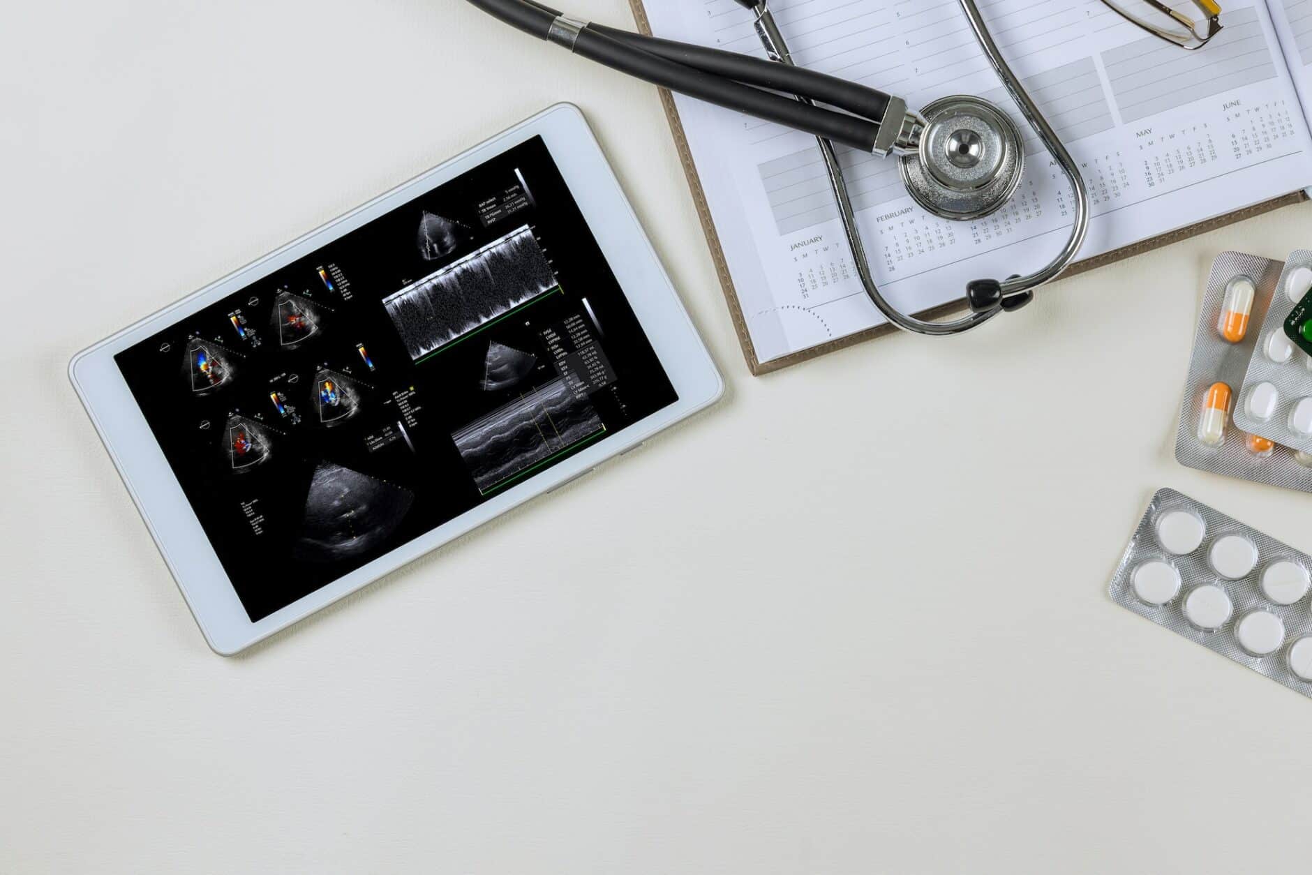
While cardiovascular medicine has had its share of breakthroughs over the past several decades, few have been as impactful on patient care as the introduction of ultrasound. Ultrasound technology gives physicians a powerful diagnostic tool that’s non-invasive, safe, and versatile.
However, to master the art of imaging and echocardiography, it’s important first to understand the underlying science that drives ultrasound technology – ultrasound physics. Keep reading to learn more about how a better understanding of ultrasound physics can help improve your cardiac imaging.
The Basics Of Ultrasound Physics
Ultrasound physics studies the principles and concepts governing the generation, propagation, interaction, and detection of high-frequency sound waves used in medical imaging, communication, navigation, and industrial processes.
Ultrasound refers to sound waves with frequencies above the upper limit of human hearing, about 20 kHz. These high-frequency sound waves are used in various industries and applications, including medical imaging and diagnostics.
Sound Waves And Their Properties
Sound waves are central to generating echocardiographic images. These mechanical waves are generated by vibrations that cause pressure variations in the propagation medium, for example, air or tissue.
The generation of ultrasound waves is based on a phenomenon known as piezoelectricity, which is the ability of certain materials to generate electrical charges when subjected to mechanical stress.
Their properties primarily determine the behavior of sound waves as longitudinal waves. This means that particles oscillate parallel to the wave’s propagation direction, displacing the surrounding particles one after the other.
Understanding these fundamental principles underlying the behavior of sound waves is essential for physicians working with echocardiography. It enables them to adjust the device parameters to achieve optimal image quality while minimizing potential errors or artifacts.
Transducers And Their Functions
As a key component in ultrasound imaging, transducers perform two essential functions. They convert the electrical energy the transmitter supplies into sound pulses and receive the reflected echoes.
Ultrasound transducers are available in various designs and play a critical role in meeting multiple imaging needs for medical professionals like cardiologists.
Frequency And Wavelength
Frequency refers to the number of cycles a sound wave completes per unit of time, typically measured in Hertz (Hz), while wavelength is the distance between two consecutive points on a wave with similar properties. These two terms have an inverse relationship, i.e., as frequency increases, wavelength decreases, and vice versa.
Higher-frequency ultrasound waves provide better resolution and allow more detailed images of the structure and function of the heart.
On the other hand, ultrasound waves with lower frequencies have longer wavelengths, which can penetrate deeper into the tissue.
High-frequency transducers are preferred for imaging superficial structures or assessing fine anatomical details. While low-frequency transducers are more suitable for imaging deeper structures, such as the heart, in patients with a larger body habitus or increased chest wall thickness.
Ultrasound In Clinical Settings
In clinical settings, ultrasound plays a significant role in the diagnostic process, as it allows healthcare providers to visualize internal organs, tissues, and other structures within the body without invasive procedures.
Ultrasound technology is regularly used in clinical settings for a wide variety of exams, including:
- Abdominal examinations
- Cardiac assessments
- Maternity and gynecological examinations
- Urological and cerebrovascular evaluations
- Breast examinations
- Assessing small tissue pieces
- Pediatric examinations
- Intraoperative guidance
While some ultrasound examinations involve using a transducer inside the body, such as a transesophageal echocardiogram, ultrasound is typically non-invasive.
Echocardiography: The Art Of Cardiac Imaging
Echocardiograms, also known as cardiac ultrasounds or simply “echo,” are noninvasive diagnostic tools that use ultrasound technology to provide detailed information about the heart and its surrounding structures.
Cardiologists first adopted ultrasound technology for diagnostic purposes in the 1960s. The introduction of cardiac imaging allowed medical professionals to visualize the heart’s structure and function without requiring an invasive procedure.
Over time, emergency physicians also began using echocardiography as part of their point-of-care ultrasound applications, spanning from head to toe and including procedural guidance.
Cardiac ultrasound can be used for a variety of indications, including:
- Significant EKG changes
- Assessing left ventricular function
- Chest pain or palpitations
- Dizziness
- Shortness of breath
- Hypotension
- New heart murmurs
- Cardiac arrest (assessing for cardiac standstill)
Types Of Echocardiograms
Several different types of echocardiograms can be used to create images of the heart, each with its own unique purpose to provide useful information about the heart. These include:
- Transthoracic Echocardiogram (TTE): The most common type of echocardiogram, TTE is a noninvasive procedure that occurs entirely outside the body. During this test, a technician applies gel to the patient’s chest and uses a handheld transducer to scan the heart. TTE provides a standard assessment of the heart’s structure, function, and blood flow.
- Transesophageal Echocardiogram (TEE) – A TEE may be performed if a standard TTE does not provide sufficient detail. This test involves inserting a special ultrasound probe into the patient’s mouth and down their esophagus after sedation. As the esophagus and heart are in close proximity, TEE can produce clearer images without interference from skin, muscle, or bone.
- Stress Echocardiogram – This test is conducted before and after exercise to evaluate how the heart responds to physical activity or stress. It is particularly useful for diagnosing coronary artery disease. If a patient cannot exercise, medication may be administered to simulate the effects of physical exertion on the heart.
- Fetal Echocardiogram – Performed during pregnancy, this noninvasive test allows healthcare providers to examine the unborn baby’s heart. By moving an ultrasound wand over the pregnant person’s belly, doctors can detect potential congenital heart defects and monitor the baby’s heart development.
- 3D Echocardiogram – Available in some medical centers and hospitals, this advanced technology provides three-dimensional images of the heart, offering more detailed information about the heart valves, replacement heart valves, and the left ventricle.
- Contrast-Enhanced Echo – A special type of echocardiography that can differentiate blood flow in different heart parts by injecting a contrast agent into a vein before imaging. This technique improved echocardiographic resolution and helps provide real-time assessment of blood flow.
- M-Mode Echocardiogram: This simple form of echocardiography produces a tracing-like image of the heart structures, enabling measurement of the heart’s chambers, size, and wall thickness.
The Doppler Effect & Echocardiography
The Doppler effect is a fundamental principle in ultrasound technology that gives medical professionals insights into blood flow velocities and direction within the heart and blood vessels. It’s used to measure blood velocity, flow direction, and Doppler shift using color Doppler imaging.
Velocity Measurement
When assessing a patient’s blood flow, one of the primary concerns is the velocity, or speed, of the blood flow. This measurement is an important marker in the diagnosis of various heart diseases.
Echocardiograms measure blood flow velocity by analyzing the Doppler shift frequency and applying a formula that considers known values such as the speed of sound in the blood and the intersection angles between the ultrasound beams and the bloodstream.
Flow Direction
Flow direction is another parameter assessed in Doppler echocardiography that helps diagnose heart disease. Direction is determined by whether the Doppler frequency shift (DFS) is positive or negative.
When erythrocytes (red blood cells) flow toward a transducer, they compress the sound waves and increase frequency, resulting in a positive DFS. On the other hand, when erythrocytes move away from the transducer, they stretch the sound waves and decrease their frequency, resulting in negative DFS.
For example, when assessing the speed of blood flow through a narrowed valve opening, known as stenosis, the physician can determine whether blood is flowing too slowly or too quickly through the opening, depending on whether there’s turbulent flow with high-frequency shifts or laminar flow with low-frequency shifts. This allows the echo reader to then determine the severity of valve stenosis.
Doppler Shift
Doppler shift refers to the change in frequency of sound waves that occurs when there’s relative motion between the source of the wave and its receiver.
In echocardiography, Doppler shift measures blood velocity and flow direction in the heart. By emitting an ultrasound wave reflected by moving blood cells, we can detect frequency changes related to the speed at which these cells move toward or away from us.
This information provides valuable insight into heart function and helps diagnose various heart diseases.
Color Doppler Imaging
Color Doppler Imaging displays color-coded maps showing the speed and direction of blood flow in the heart in addition to traditional grayscale images.
Medical professionals rely heavily on color Doppler ultrasound technology because it helps identify blockages or narrowing in arteries leading to certain body parts. Additionally, color Doppler is used to detect narrowings or backflow (regurgitation) of heart valves.
When screening for diseases of the carotid artery that affect the blood supply to the brain, strokes can be significantly reduced when detected early through color Doppler ultrasound screening, saving lives.
The Primary Applications of Echocardiography
The primary applications for echocardiography are vast, spanning the diagnosis and management of various cardiac conditions. Using ultrasound imaging, medical professionals can help diagnose and monitor various heart diseases, guide procedures, and assess heart function.
Assessing Heart Function
The primary use for echocardiography is to provide doctors with a non-invasive assessment of a patient’s heart function. Some of the key pieces of data that an echo can provide insight on are:
- Ejection fraction (EF) is the percentage of blood the left ventricle ejects with each heartbeat. A normal EF range is between 50% and 70%. An EF below 40% indicates cardiac dysfunction that can lead to heart failure.
- Fractional Shortening (FS) measures the contraction of the left ventricle. FS Values should range from 28% to 45%.
- Wall motion abnormalities indicate changes in the motion patterns or movement of the cardiac wall segments, which may indicate myocardial infarction.
- Diastolic Function measures how efficiently the ventricles fill with blood during the relaxation phase. A lack of relaxation can cause diastolic dysfunction, which leads to heart failure.
- Valvular Function evaluates problems with the valve leaflets, annulus, chords, and coaptation that cause narrowing or insufficiency of valve flow.
Heart Disease Diagnoses
Echocardiography is the most commonly used imaging technique for diagnosing cardiovascular disease for a few key reasons:
- Echocardiography allows for non-invasive diagnosis of various pathological heart conditions such as myocardial infarction, valvular disease, and cardiomyopathies.
- Real-time imaging allows the detection of structural abnormalities such as chamber enlargement, valve thickening, and wall motion abnormalities.
- Doppler techniques allow the assessment of hemodynamic changes such as blood flow velocity and direction, which can help diagnose conditions such as atrial septal defects and aortic stenosis.
- Stress echocardiography can assess coronary artery disease by measuring changes in cardiac function under stress.
- 3D echocardiography allows better visualization of complex cardiac structures such as congenital heart defects or prosthetic valves.
Monitoring Heart Conditions
Echocardiography allows physicians to access detailed information about their patients’ hearts through regular monitoring without exposing them to additional risks. Some key uses are:
- Echocardiography can measure the ejection fraction, an important parameter for assessing left ventricular function. Changes in ejection fraction may indicate worsening heart failure or other cardiac problems.
- It can detect abnormalities of the heart valves, such as stenosis or regurgitation. Monitoring these abnormalities over time can help guide treatment decisions.
- Echocardiography can reveal changes in the size and thickness of the heart chambers and walls. This information can help diagnose and monitor conditions such as hypertrophic cardiomyopathy.
- Patients with pacemakers, defibrillators, or other implanted cardiac devices may need regular echocardiograms to ensure the devices are working properly and not causing complications.
- Echocardiography can monitor how patients respond to medications or other treatments for heart disease. Repeated echocardiograms can show if a treatment is positively affecting the heart.
Guiding Procedures
Ultrasound can also be useful to guide physicians during various medical procedures to reduce risk and improve patient outcomes. A few key procedures that use cardiac ultrasound technology are:
- Image guidance during cardiac catheterization.
- Guidance of needle placement during pericardiocentesis, epicardial biopsy, or drainage of cardiac fluid.
- Transthoracic echocardiography (TTE) and transesophageal echocardiography (TEE) to guide transcatheter valve replacement or repair.
- Use of ultrasound images to assist in the implantation or removal of pacemakers.
- Guidance of fetal interventions and surgeries.
Future Advancements And Limitations In Ultrasound Technology
Ultrasound technology has come a long way since its inception, with continuous advancements leading to improved image quality, portability, and functionality. However, limitations still need to be addressed as the field continues to evolve.
Recent Advancements In Ultrasound Technology
Recent advances in ultrasound technology have directly contributed to improved patient care and more accurate diagnosis, allowing for improved treatment of various medical conditions.
Some of the more significant advances we’ve seen in recent years include:
- Volumetric Ultrasound Technology allows for creating images with greater depth and detail than traditional sonograms. Volumetric ultrasounds have proven to be particularly useful in identifying tumors, analyzing cardiac function, and examining various other internal structures.
- Another recent advancement is 4D sonography technology which adds the dimension of time to ultrasound exams, allowing sonographers to create 3D images in real-time. This provides a more comprehensive view of internal organs and structures, allowing for better visualization and diagnosis.
- Contrast-Enhanced Ultrasound (CEUS) technology uses contrast agents to improve the clarity of images and more easily detect abnormalities. This has demonstrated effectiveness in tumor detection, a task typically reserved for more advanced tests like CT or MRI scans
- Portable and hand-held ultrasound devices are now available in various settings, including emergency rooms, operating rooms, and ambulances.
- Elastography measures tissue elasticity to diagnose liver fibrosis, breast masses, and prostate cancer.
- Artificial Intelligence (AI) software can help automate image interpretation and analysis for more efficient diagnoses.
- HIFU (High-Intensity Focused Ultrasound) Therapy uses concentrated ultrasound waves to destroy cancerous tissue without surgery.
Technical Limitations
Challenges related to operator dependency, patient factors, and imaging depth and resolution must be addressed to fully harness these innovations’ potential.
Once these limitations are overcome, ultrasound technology can continue to evolve and play an increasingly vital role in diagnosing and managing cardiovascular diseases. Some of the main technical limitations include:
- Operator Dependency: Inadequate training or technique can lead to suboptimal image quality and diagnostic errors. Ongoing education, training, and standardization of protocols can help overcome this challenge.
- Acoustic Windows and Patient Factors: The quality of ultrasound images can be compromised by patient factors such as obesity, lung disease, or surgical dressings, which can limit the available acoustic windows for imaging. To address this limitation, new transducer technologies or imaging techniques may need to be developed to overcome these obstacles and provide clear, high-quality images.
- Imaging Depth and Resolution: Ultrasound technology currently faces a trade-off between imaging depth and resolution, as higher frequencies provide better resolution but reduced penetration depth. Future advances in transducer technology and signal processing may help find a better balance that provides both high-resolution imaging and adequate penetration depth for various clinical applications.
Ready For A Deeper Dive Into Echocardiography?
If you want to improve your understanding of cardiac imaging and echocardiography, you’ve come to the right place. We’ve put together a comprehensive online course for medical professionals that provides in-depth instruction on everything you’ll need to know about Point-of-Care Echocardiography.
Our two-hour course covers general topics such as basic cardiac anatomy, how to obtain images, and more in-depth discussions on calculating the ejection fraction, left ventricular function, and assessing wall motion abnormalities.
In this course, you won’t just memorize images but, instead, learn how to assess the heart from any view and scrutinize normal and abnormal findings.
Point Of Care Echo is available in both CME and Non-CME formats, making it a great course for emergency department providers, intensive care unit providers, most hospitalists, all medical students, nurse practitioners, physician assistants, and emergency medical technicians.
Click here to create your free account with ECGEDU.com today!


