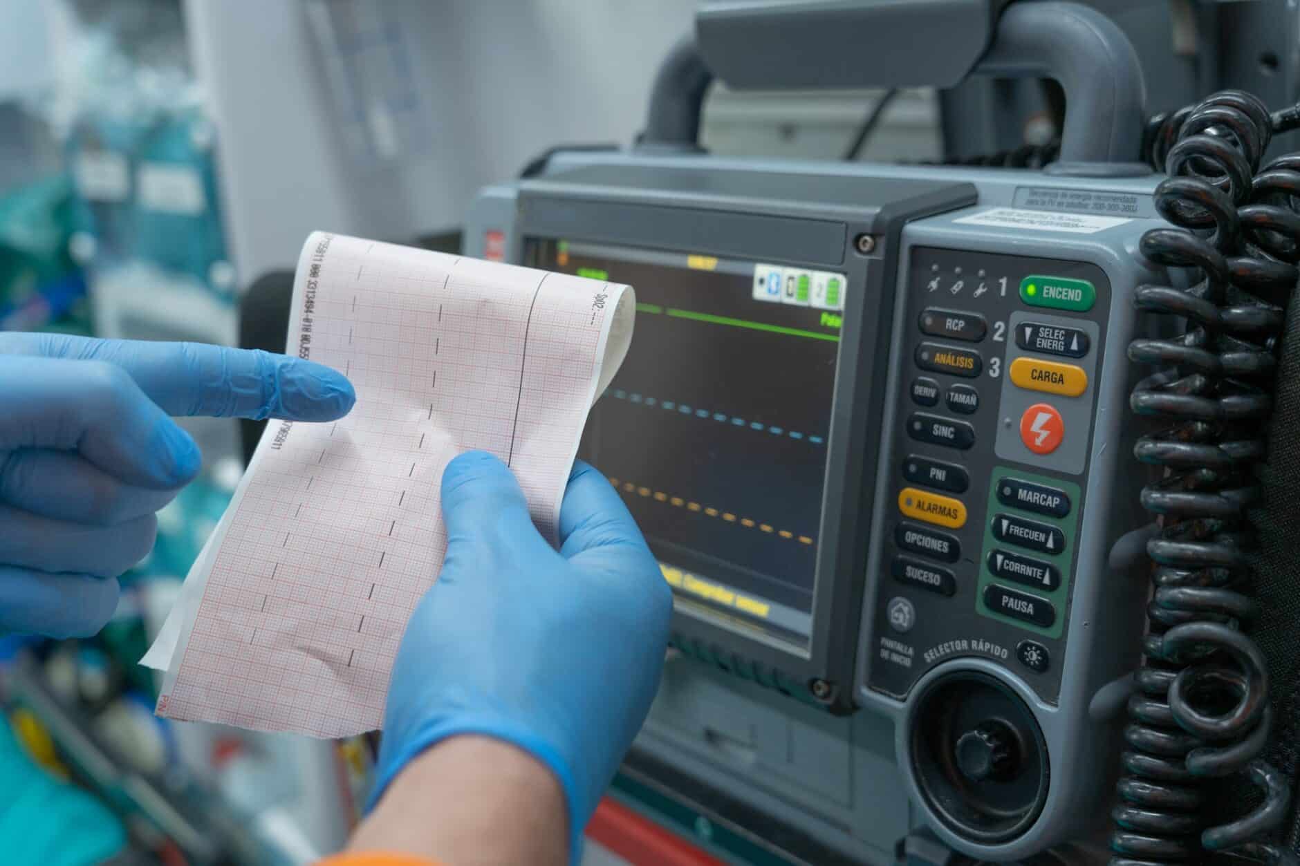
As every physician one day learns, “false alarm” rhythms can show up on an ECG reading that can closely resemble life-threatening rhythms, such as an ST-segment elevation myocardial infarction (STEMI). Advanced care providers need to quickly differentiate these rhythms, known as ECG mimics, from their more serious counterparts to avoid unnecessary treatment or allocation of hospital personnel.
Keep reading to learn more about common ECG mimics, identification techniques, and strategies and tactics to prevent misdiagnosis.
Understanding ECG Mimics
ECG mimics are non-cardiac abnormalities that can mimic cardiac conditions on an electrocardiogram. They may closely resemble other serious cardiac conditions such as acute coronary syndrome (ACS), myocardial infarction (MI), ST-segment elevation myocardial infarction (STEMI), and various arrhythmias.
These similarities can pose a challenge to medical personnel that may be less experienced in interpreting ECG results, increasing the risk of misdiagnosis and inappropriate treatment.
Common ECG Mimics And How To Identify Them
Because ECG mimics resemble serious cardiac events, learning to identify common mimics and their signs can help avoid unnecessary misdiagnosis and even make the difference between life and death. Here are a few of the most common mimics and ways to identify them.
Early Repolarization
Early repolarization is a common, benign ECG finding observed in healthy individuals, particularly young adults, and athletes. It’s characterized by J-point elevation, often accompanied by notching or slurring of the terminal QRS complex.
However, this normal variant can sometimes be mistaken for ST-segment elevation myocardial infarction (STEMI), a critical condition requiring immediate medical intervention.
Differentiating early repolarization from STEMI involves careful analysis of the ST-segment morphology, reciprocal changes, distribution of ST-segment elevation, additional ECG findings, and the clinical context. Accurate recognition of early repolarization is important to avoid unnecessary interventions and ensure appropriate patient care.
Left Ventricular Hypertrophy
Left ventricular hypertrophy (LVH) is a common finding in clinical practice, with hypertension being the most common cause. LVH is recognized as a cause of many STEMI (ST-Elevation Myocardial Infarction) mimics, and it can be challenging to determine whether ST changes are present.
If left undiagnosed or misdiagnosed, LVH can mimic an acute MI, leading to inappropriate treatment and management.
One way to detect LVH is with an echocardiogram, which measures the thickness of the heart muscle walls. When LVH occurs without underlying disease-known as mimic hypertrophic cardiomyopathy-other tests may be needed in addition to treatment for hypertension.
Pericarditis
Pericarditis is a common cause of chest pain that can be easily misdiagnosed if ECG changes aren’t recognized. The classic ECG changes associated with pericarditis develop in four progressive stages, beginning with diffuse ST-segment elevation and PR-segment depression in all leads, which may resemble myocardial infarction.
In addition, pericarditis may present with subtle signs and symptoms that may go unnoticed without adequate investigation, resulting in delayed diagnosis and treatment.
To differentiate pericarditis from a true cardiac event, look for concave-upward ST-segment elevation in multiple leads, reciprocal PR-segment elevation in aVR, and consider the patient’s symptoms and clinical context.
Electrolyte Imbalances
Electrolyte imbalances can cause significant ECG abnormalities mimicking a broad spectrum of cardiac diseases, leading to misdiagnosis and inappropriate treatment. Hyperkalemia, hypokalemia, hypercalcemia, and hypocalcemia are the most common electrolyte disturbances that affect ECG signals.
For example, hyperkalemia may manifest as peaked T waves or QRS complex widening, while hypokalemia may result in ST segment depression or a prolonged QT interval. In addition, multiple electrolyte disturbances may occur simultaneously, further complicating the diagnosis.
To identify hyperkalemia as the cause of these ECG findings, consider the patient’s clinical presentation, review laboratory results for potassium levels, and assess for other conditions that may cause similar ECG changes.
ECG Artifacts
Artifacts on an ECG can lead to misinterpretation of abnormal rhythms or patterns, potentially resulting in incorrect diagnoses and inappropriate treatments. Common artifacts that can interfere with ECG measurements include baseline wander, muscle tremors, and interference from nearby electrical equipment.
Proper skin preparation, secure electrode attachment, and appropriate lead placement are important to obtain accurate ECG readings. Being mindful of the patient’s and environmental movements can help identify potential sources of artifacts and improve the overall quality of the ECG tracing.
Medication Side Effects
There are also several prescription medications that can cause mimics to show up on an ECG. For example, Digoxin or antiarrhythmic drugs can cause arrhythmias that resemble other ECG patterns.
This is one of the reasons it’s critical to obtain a comprehensive medical history, including information about current medications and any adverse effects in the past on a patient. Clinicians must also be alert to drug-drug interactions, which can affect cardiac function and lead to misleading ECG findings.
Strategies For Avoiding Misdiagnosis
Misdiagnosis can have serious implications for patient care, leading to unnecessary treatments, delayed interventions, higher healthcare costs, and increased patient anxiety. It’s important for healthcare professionals to implement strategies to minimize the risk of misdiagnosis and ensure proper clinical decision-making.
Conducting A Comprehensive Patient History And Physical Examination
An essential step in accurately interpreting an ECG mimic is a comprehensive patient history and physical examination. This allows the healthcare professional to gather important information about the patient’s medical history, symptoms, risk factors, medications, and family history that can assist in making the correct diagnosis.
During the physical examination of patients with cardiac symptoms consistent with potential ECG mimicry conditions such as left ventricular hypertrophy (LVH), the physician can look for signs such as enlarged precordial motion or extra heart sounds unique to LVH patients.
A full-body exam also helps detect electrolyte imbalances resulting from underlying medical problems such as renal failure.
Perform Repeat ECGs And Supplemental Tests
If the initial ECG findings are inconclusive, unclear, or suggestive of artifact, you will typically perform a repeat ECG. This can help confirm the initial findings or reveal any changes in the patient’s cardiac condition. Additionally, repeat ECGs can help detect transient abnormalities or correct technical problems that may have affected the initial ECG.
Incorporating other diagnostic tests can help confirm ECG findings and provide a more complete picture of the patient’s cardiac condition. These tests include cardiac imaging or blood tests.
Cardiac imaging tests, such as computed tomography (CT), echocardiography, angiography, magnetic resonance imaging (MRI), and nuclear imaging, can provide detailed information about the structure and function of the heart and its blood vessels and help confirm or rule out certain diagnoses. Blood tests, including cardiac biomarkers such as troponin and creatine kinase-MB (CK-MB), can help identify the presence of myocardial damage and provide additional information to support ECG findings.
Seeking Consultation From Cardiac Specialists
Consulting with a cardiac specialist is also an excellent way to avoid ECG mimic misdiagnosis. Cardiac specialists bring a unique and valuable perspective to diagnosing and treating cardiovascular disease, including ECG abnormalities.
Experts recommend prompt consultation with a specialist when a patient’s clinical presentation is inconsistent with initial diagnostic testing or when there’s uncertainty in interpreting test results.
Consulting cardiac specialists not only provides expert advice but can also help avoid common pitfalls, such as premature hospital discharge or the initiation of inappropriate treatment due to misinterpretation of ECG findings.
Technological Advancements In ECG Interpretation
Advances in healthcare technology have led to the development of sophisticated algorithms and decision support systems capable of analyzing and interpreting Electrocardiogram (ECG) monitoring data. These systems use a combination of artificial intelligence (AI) and machine learning (ML) to recognize patterns in the ECG data and provide clinicians with real-time insights into the patient’s heart rate, rhythm, and other vital signs.
By leveraging the power of AI, these systems can detect even subtle changes in the patient’s ECG data and alert clinicians to potential problems before they become serious.
In addition to providing real-time insights into the patient’s cardiac health, these systems can be used to monitor trends over time. By tracking changes in the ECG data, clinicians can detect changes in the patient’s heart rate, rhythm, and other vital signs that may indicate a developing condition or potential risk. This can allow clinicians to intervene and provide treatment before the condition becomes more serious.
A study published in the Journal of Cardiovascular Electrophysiology found that using a decision support system improved physicians’ accuracy in detecting abnormal ECGs by 15%. In another study, researchers from MIT developed an algorithm that could predict whether a person was at risk for sudden cardiac arrest by analyzing ECG results.
As machines become better at recognizing patterns in large data sets and detecting anomalies more accurately than humans ever could on their own, it’ll become increasingly important to incorporate ML and AI into clinical decision-making processes.
Ten Ways To Identify An ECG Mimic
ECG interpretation is just as much of an art as a science, so the best way to ensure accurate identification of ECG mimics is through practice, repetition, and experience. However, when in doubt, here are ten ways to more accurately interpret a mimic from the real thing.
- Review the patient’s medical history thoroughly, paying particular attention to cardiac or neurologic symptoms.
- Check for technical problems with ECG measurement, such as poor electrode attachment or interference from external devices.
- Monitor the patient’s electrolyte levels, as imbalances can lead to ECG changes that mimic other conditions.
- Rule out medication side effects by reviewing the patient’s current medications and their potential impact on the heart.
- Identify heart rhythm disturbances, including tachycardia, bradycardia, and atrial fibrillation.
- Look for signs of left ventricular hypertrophy or thickening of the muscle of the left ventricle.
- Look for common signs of myocardial infarction, such as pericarditis or pulmonary embolism.
- Watch for normal variants that may appear on an ECG but do not indicate a pathologic condition.
- Consider additional testing, including repeat ECGs and imaging studies such as echocardiography, to investigate any other abnormalities further.
- If needed, consult with a cardiac specialist to ensure optimal treatment and therapy decisions.
Want To Learn More? Sign Up For One Of Our ECG Interpretation Courses Today!
As a medical professional, you understand the importance accurate ECG interpretation plays in diagnosing and managing cardiac conditions. With constant advancements in technology and new insights emerging in the field, staying up-to-date with the latest knowledge and best practices is essential for delivering the highest standard of patient care.
ECGEDU.com offers a wide range of self-paced video courses that make electrocardiogram and arrhythmia interpretation simple and understandable. Our ECG interpretation courses are personal and interactive and include hours of video content, ECG practice tests, helpful ECG resources, and even Category 1 AMA or AOA CME Credits.
Click here to register for your free account on ECGEDU.com and start improving your ECG interpretation skills today.


