If you’ve ever looked at an ECG reading, you may notice that readers will occasionally mention an rSR’ pattern in their interpretation. While this may sound like cause for alarm, most of the time, it’s actually a benign finding.
Keep reading to get an in-depth overview of what an rSR prime (rSR’) pattern is, as well as some of the common and uncommon etiologies associated with it. We will also examine the criteria for diagnosing Right Bundle Branch Block (RBBB), explore its possible causes, and outline its clinical implications for patient health management.
First, The QRS Complex
Before we dive into the rSR’ pattern, it’s important to give an overview of the QRS complex. The QRS complex represents ventricular depolarization, which is the process by which the electrical impulse spreads through the ventricles, initiating their contraction. This complex is key to understanding the heart’s function, as it indicates the beginning of systole and ventricular contraction.
Each wave within the QRS complex specifies a specific area and direction of the electrical activity of the heart. For example, if you look at lead two, the small Q wave corresponds to the depolarization of the interventricular septum, whereas the R waves, the largest of the three waves, reflect the depolarization of the main mass of the ventricles.
Finally, the small S wave represents the final depolarization occurring in the Purkinje fiber near the heart’s apex. The normal duration of the QRS complex is between 0.08 and 0.10 seconds, with a duration greater than 0.12 seconds considered abnormal.

Start Your Membership Today
We make electrocardiogram interpretation simple and understandable. The videos are interactive, and have detailed, easy to follow illustrations.
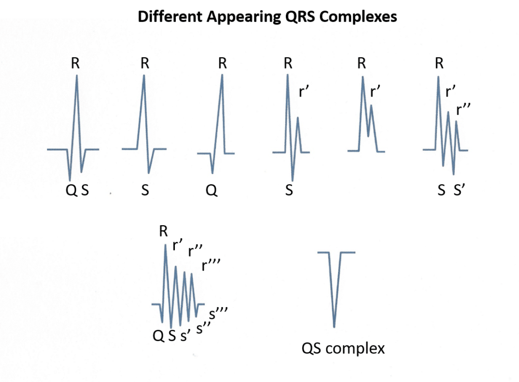
The components of the QRS Complex are:
- Q-wave: The first downward deflection before the R-wave
- R-wave: The first upward deflection of a QRS complex
- S-wave: The first downward deflection after the R-wave
- R prime (R’): The second upward deflection of a QRS complex
- S prime (S’): The second downward deflection after the R-wave
- Subsequent upward deflections: R double prime (R”), R triple prime (R”’),…
- Subsequent downward deflections: S double prime (S”), S triple prime (S”’),…
What Is An RSR Prime (rSR’) Pattern?
In its simplest terms, an rSR prime (rSR’) pattern is nothing more than a QRS complex with an upward deflection (R wave), followed by a downward deflection (S wave), and followed by a second upward deflection (R prime).
Although this pattern may be seen in any ECG lead, it is only mentioned when seen in leads V1 and/or V2. When seen in other leads, it is usually insignificant. Additionally, when ECG readers point out an rSR’ it means that the QRS interval is normal (0.08-0.10 seconds) or only mildly prolonged (0.11 seconds).
In some cases, you may also see a small “r” or a large “R” to denote which upward deflection is bigger. For example, rSR’ indicates a small R- wave, followed by an S- wave, followed by a large R wave. Similarly, RSr’ indicates a large R wave, followed by an S wave, followed by a small R wave.
Definition And Characteristics Of RSR’ Pattern
The RSR Prime (rSR’) pattern represents a delay in activation within the basal part of the right ventricle (RV), which can be observed through distinct variations in QRS complex morphology.
Several types of rSR’ patterns exist, such as incomplete or complete right bundle branch block (RBBB) patterns. Incomplete RBBB often manifests as an rSr’ pattern in leads V1-3 with QRS duration less than 120ms—a common finding among children without clinical significance.
Conversely, complete RBBBs exhibit rsr’, rsR’, or rSR’ configurations in ECG leads V1-V2 with QRS durations exceeding 120ms; these conditions warrant further investigation from healthcare providers to determine proper diagnosis and treatment plans for affected individuals.
Types Of RSR’ Patterns
Multiple different RSR’ patterns can potentially be observed, each with distinct characteristics and potential underlying causes. Medical professionals need to recognize these various patterns to diagnose and treat their patients appropriately. Some common types of RSR’ patterns include:
- Right Bundle Branch Block (RBBB): This pattern presents as a prolonged QRS complex duration, typically greater than 120 ms, accompanied by a slurred terminal R wave in precordial leads V1-V2 and a wide, deep S wave in lateral leads I, V6.
- Brugada Syndrome: An rSr’ pattern in leads V1-V2 can indicate this genetic disorder causing sudden cardiac death due to ventricular arrhythmias. The ECG findings include ST-segment elevation in right precordial leads V1-V3 with a characteristic coved-type morphology.
- Incomplete RBBB (IRBBB): An rSR’ pattern seen with an otherwise normal or only slightly prolonged QRS complex duration may represent a benign variant or transient condition.
- Arrhythmogenic Right Ventricular Cardiomyopathy (ARVC): A potentially life-threatening condition characterized by fibrofatty replacement of the right ventricular myocardium that can manifest as an rSR’ pattern on ECG.
- Precordial Lead Misplacement: High placement of the precordial lead can mimic the appearance of an rSR’ pattern in leads V1-V2; Ensuring correct lead positioning is crucial for accurate ECG interpretation.
Being familiar with these different types of RSR’ patterns in ECG will allow doctors to identify any underlying conditions and guide patient management effectively and quickly.
Identifying An RSR’ Pattern In ECG Results
To accurately identify an rSR’ pattern in ECG results, healthcare professionals must consider various factors, including the patient’s age, medical history, and any existing heart conditions. In young people and athletes, an rSR’ pattern with a QRS duration of less than 120 ms may be due to an incomplete right bundle branch block (IRBBB) or a normal electrophysiological variant. However, it is essential to recognize and diagnose serious pathological conditions that may also present with this pattern.
Interpreting ECG Results
When analyzing the QRS complex, physicians should pay close attention to its duration, morphology, and axis.
A wide QRS complex (>120ms) with an M-shaped pattern in V1 and a wide, slurred S wave in lateral leads may indicate Right Bundle Branch Block (RBBB). However, other non-cardiac factors can also contribute to an rSR’ pattern on ECG, including normal variants such as juvenile RBBB or incomplete RBBB.
Clinical events like acute pulmonary embolism or myocardial infarction can also lead to transient changes in the ECG.
Identifying Potential Underlying Causes
When diagnosing the RSR’ pattern in ECG, it is essential to identify potential underlying causes. While an rSR’ pattern in leads V1-V2 on electrocardiogram is fairly commonplace in clinical cardiology, that doesn’t always mean there’s not a more serious underlying cause.
For example, serious medical conditions such as pulmonary embolism or COPD may cause an RSR’ pattern. Additionally, congenital heart defects such as tetralogy of Fallot or an atrial septal defect can also present with this ECG pattern.
It’s important to remember that each patient is an individual and that what may be normal for one might be life-threatening for another.
Medical Conditions Associated With RSR’ Pattern
The rSR’ pattern in ECG is associated with several medical conditions, including:
- Right Bundle Branch Block (RBBB): This is the most common cause of an rSR’ pattern in an ECG. It occurs when there is a delay or blockage in the electrical conduction through the right bundle branch of the heart.
- Brugada Syndrome: This inherited condition affects the heart’s electrical system and can lead to sudden cardiac arrest or death. The rSR’ pattern in leads V1-V3 usually has a distinct pattern cluing you in to Brugada syndrome.
- Malignant ventricular conditions: In some cases, an rSR’ pattern in ECG can indicate underlying malignant ventricular conditions such as arrhythmogenic right ventricular dysplasia (ARVD) or hypertrophic cardiomyopathy.
- Other heart diseases: The rSR’ pattern may also be present in other heart diseases, including myocardial infarction, pulmonary embolism, and dilated cardiomyopathy.
It is essential for doctors to identify and diagnose any underlying medical conditions associated with the rSR’ pattern in ECG to provide proper treatment and management strategies for their patients.
What Is The Significance of an rSR’ in Leads V1 or V2?
As mentioned above, most of the time an ECG reader mentions an rSR’ pattern on the ECG, the QRS complex is narrow or only mildly prolonged. Most of the time, this finding is benign and is caused by abnormally high lead placement of V1 and V2. This is a normal variant seen due to normal depolarization of the intraventricular septum or an incomplete right bundle branch block.
Additionally, this pattern can be seen in athletes or people with sunken chests (pectus excavatum). In some cases, the rSR’ is significant. Pathological conditions causing this include Brugada Syndrome, right ventricular enlargement, right ventricular hypertrophy, or abnormal conduction through a bypass tract with preexcitation or Wolff Parkinson White (WPW).
Hopefully, the ECG interpreter can tease these diagnoses out in these cases and be more specific with their reading.
Monitoring And Management Strategies
In some patients, ongoing monitoring may be necessary to determine whether there is a health risk or the finding is benign. In these situations, a physician might choose one of the following strategies.
- Regular electrocardiograms (ECG) to track changes in the RSR’ pattern and evaluate any new arrhythmias or conduction abnormalities.
- Echocardiography to assess the heart’s overall health and detect any structural abnormalities.
- Holter monitor or event recorder to capture any transient arrhythmias that may not be detected on a standard ECG.
- Treatment of underlying medical conditions, such as hypertension or coronary artery disease, to reduce the risk of further cardiac complications.
- Close follow-up with a cardiologist for ongoing evaluation and management of any identified issues.
Ready To Learn More About ECG Interpretation?
If you are a physician, medical student, or medical professional, there is no downside to improving your ECG interpretation skills.
Our self-paced, interactive videos make electrocardiogram interpretation simple and understandable, and the courses are designed to give a one-on-one feel to learning how to read an ECG.
Our courses are designed for learners at any level, so whether you’re a nurse practitioner, a medical student, or even a practicing physician in need of an ECG CME credit, we have something for you on ECGEDU. Click here to create your account today.
References:
(1) https://onlinelibrary.wiley.com/doi/epdf/10.1111/anec.12241
Baranchuk, A., Enriquez, A., García-Niebla, J., Bayés-Genís, A., Villuendas, R. and Bayés de Luna, A. (2015), Differential Diagnosis of rSr’ Pattern in Leads V1-V2. Comprehensive Review and Proposed Algorithm. Ann Noninvasive Electrocardiol, 20: 7-17. https://doi.org/10.1111/anec.12241

